News • Rhabdomyosarcoma
Muscle cancer - or is it?
Pinpointing the cell type that turns malignant in rhabdomyosarcoma opens the path for better treatments.
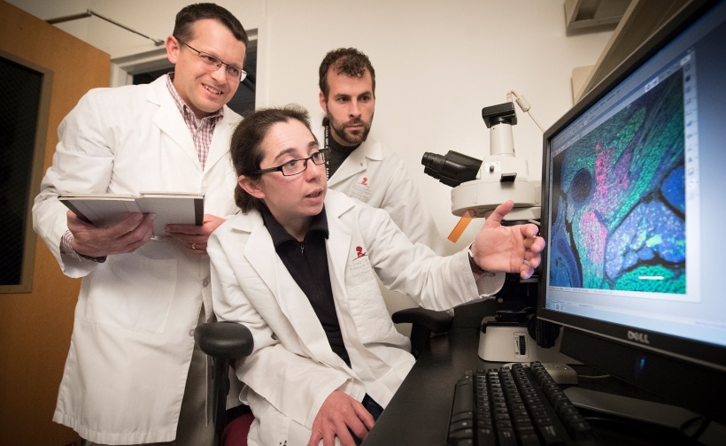
St. Jude Children’s Research Hospital oncologists have discovered the cell type that gives rise to rhabdomyosarcoma, the most prevalent soft tissue cancer in children. Previously, scientists thought the cancer arose from immature muscle cells, because the tumor resembled muscle under the microscope. However, the St. Jude researchers discovered the cancer arises from immature progenitors that would normally develop into cells lining blood vessels. The researchers, led by Mark Hatley, M.D., Ph.D., of the Department of Oncology, published their findings in the scientific journal Cancer Cell. Hatley said understanding the cell of origin will bring badly needed insights to aid diagnosis and treatment of rhabdomyosarcoma. “We are still using the same chemotherapy that was in use 46 years ago, with the same outcomes,” Hatley said. “A better understanding of the machinery of rhabdomyosarcoma could enable entirely new treatment approaches.
These tumors were not driven by muscle cells at all, so we decided to zero in on the biological machinery
Mark Hatley
“While these tumors appear to be muscle cells under the microscope, and clinicians had thought that they arose from muscle progenitor cells, that didn’t explain why the tumors can occur in tissues that don’t have skeletal muscle, like bladder, prostate and liver,” he continued.
Hatley said Andrew McMahon, then at Harvard University, had genetically engineered a mouse to have a biological switch that enabled researchers to selectively turn on a key piece of cellular machinery called the Hedgehog pathway. Abnormal activation of this pathway was known to trigger cancers. Jonathan Graff at the University of Texas Southwestern Medical Center used this model to study the role of the hedgehog pathway in fat cell development, however the animals developed head and neck tumors. Hatley, along with Rene Galindo, Eric Olson and in collaboration with Graff, determined these tumors were rhabdomyosarcoma. “These tumors were not driven by muscle cells at all, so we decided to zero in on the biological machinery to find the cell of origin in these mouse tumors,” Hatley said.
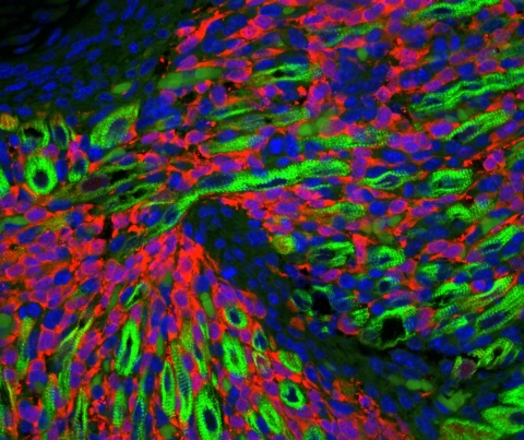
His experiments revealed the cells that became rhabdomyosarcoma were not muscle cells, but were immature cells that would mature into cells lining the inner surface of blood vessels. The blood vessels occupy the space between muscle fibers. “That was a complete surprise,” Hatley said. “We also found that the tumors developed quickly, at the time in early development that corresponds to when such tumors develop in children with the cancer.”

The finding suggested the cancer process began before birth. “Indeed, when we studied the mice at the embryonic stage, we saw the cells between the muscle fibers expanded explosively and formed tumors early in development,” Hatley said. The researchers also explored whether another key cancer-activating pathway, called KRAS, might trigger rhabdomyosarcoma. Other scientists had found evidence that KRAS could drive such tumors. However, when the researchers switched on KRAS in the preclinical models, an entirely different kind of tumor formed, called angiosarcoma. “This finding told us that the tumors in our model depended on activation of the Hedgehog pathway,” Hatley said. “But it also suggested there are likely multiple cells of origin for rhabdomyosarcoma. The different location of such rhabdomyosarcomas may depend, for example, on the cell of origin.”

Detailed studies of the tumor cells revealed that, in turning malignant, the endothelial cells were abnormally reprogramming themselves during early development to be more like muscle cells. A significant finding was that the tumor cells had characteristic biological machinery that drives development of head and neck muscles. This discovery may help explain why rhabdomyosarcomas tend to occur in the head and neck. Genetic studies of the tumor cells revealed evidence of their origin as endothelial cells. “We were able to look back in the history of these tumor cells and see that they retained genes important in endothelial cell development,” Hatley said.
A major next step in the research will be to apply these preclinical findings to patients, by analyzing their tumor cells. Such studies will aid diagnosis and treatment of rhabdomyosarcoma. “If the same mechanisms hold true in our patients’ tumors, the findings could help us determine which patients will respond better to treatment,” Hatley said. “And while the tumor model we’re now studying doesn’t present targets for new drugs, if we can discover the mechanism controlling that model, it may yield therapeutic drug targets.” The findings may also lead to drugs to prevent rhabdomyosarcoma in children with a genetic predisposition to the cancer, Hatley said.
The unexpected discovery that rhabdomyosarcoma is not really a muscle cancer may offer broader lessons for researchers seeking the cellular origin of cancers. “These findings have taught us not to make assumptions about the origins of tumors based on their appearance under the microscope or the genes that are turned on,” Hatley said. “We need to seek a detailed understanding of their developmental biology, an understanding that can guide us to new treatment strategies.”
Source: St. Jude Children's Research Hospital
09.01.2018


