US radiologists wake up to risks from high radiation doses
Report: Dot M McSherry, i.t. Communications
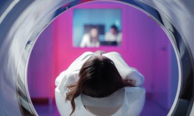
An estimated 70 million CT scans are performed annually in the USA, a threefold increase since 1993. US physicians rely on CT scans and other diagnostic imaging procedures to make accurate and speedy diagnoses and, until recently, they have not questioned the radiation dose exposure the patient receives. However, this attitude is changing, as physicians and other medical professionals realise that young and middle-aged adults have the potential to be exposed to too much radiation during their lifetime and face a higher risk of cancer.
In the US, it is not realistically possible for a physician to determine the cumulative radiation dose a patient of any age has received. Even today, radiation dose for examinations such as CT must usually be calculated manually, and the radiology staff has no time to do this. When radiation dose exposure is known, it isn’t recorded on a patient’s medical chart, whether paper or electronic.
Add to this the fragmented nature of US healthcare. Americans, a highly mobile people, may go anywhere they wish to receive medical treatment. Lack of centralised records, the vast majority of which are paper-based, places the burden of cumulative record keeping on the patient. Patients requiring complex care are referred to specialists who order more exams. Duplicative diagnostic imaging studies are frequently repeated, because even in this digital age, exams may not have been transferred in a timely manner or at all, they may not be of the quality required by the recipient hospital, or doctors may simply order procedures anew. There are few watchdogs to urge caution.
CT scanners have become ubiquitous in even the smallest US hospitals because they produce diagnostic images of excellent quality very rapidly. In most cases, their use is appropriate. But for the younger person facing a lifetime of medical care, how great is the risk of cancer in all this radiation exposure?
US radiologists and medical physicists are vocalising their concern. The well publicised Image Gently campaign started in 2008 has made huge inroads to convince radiology departments globally to ‘child size’ radiation dose and to encourage paediatricians to be conservative when ordering radiology procedures. CT vendors have developed exam protocols for children and created software algorithms to improve the quality of low-dose images.
Now concerns are being expressed about adults. Electronic patient records (EPRs) are making analysis possible. At the 2008 RSNA meeting, physicists and radiologists from Brigham and Women's Hospital in Boston, Mass. identified over 31,000 patients who had CT scans performed there or at the Dana Farber Cancer Institute in 2007. They determined that these patients had received almost 191,000 CT scans over a 22-year period. Of these, 7% had a greater lifetime risk of cancer greater than 1%, offset by the fact that 85% of this group had a serious malignancy.
In July 2009, a highly controversial article in the Archives of Internal Medicine, a peer review journal of the American Medical Association, warned that patients who undergo coronary artery calcium screening exams every five years could develop cancer from this. Physicists from the National Institutes of Health and Columbia University warned that as many as 42 in every 100,000 patients between the ages of 45 to 75 could develop radiation-induced cancer, most likely lung, breast and leukaemia. An accompanying editorial noted that there is no standard protocol for the quantification of coronary artery calcium scans.
In August 2009, the New England Journal of Medicine published a study by Dr Reza Fazel of Emory University School of Medicine in Atlanta, Georgia who used data about almost one million adults under 65 to estimate cumulative radiation doses from radiology procedures. Between 2005 and 2007, 69% of these individuals had at least one diagnostic imaging procedure. More than 80% of these exams were performed outside hospitals, in freestanding imaging centres, in clinics, and in doctors' offices. CT scans and nuclear imaging accounted for 75% of the estimated radiation dose.
The American College of Radiology (ACR) expressed its concern, stating that most of the scans were performed by non-radiologist providers that self-referred patients to their own imaging equipment. The ACR wrote that previously published studies have shown that when physicians can refer patients to their own scanners, or those in which they have a financial interest, the use of radiology procedures is greatly increased. There has been much public criticism about the increased use of diagnostic exams by cardiologists in particular.
At the RSNA 2009 a press conference at which radiology researchers from the University of Wisconsin Madison presented data that showed more than 50% of patients who undergo CT scans for suspected abdominal conditions received additional, unnecessary diagnostic procedures, was filled to capacity with journalists.
In December 2009, the Archives of Internal Medicine again published two articles that received national patient and physician attention. The U.S. National Institutes of Health and the National Cancer Institute estimated that 29,000 future cancers could be related to CT scans performed in 2007 alone, and that nearly 15,000 of those cancers could be fatal. In particular, women, children and young adults are especially susceptible, with principal author Amy Berrington de Gonzales PhD predicting that women would have two thirds of the predicted future cancers.
The second study evaluated radiation dose exposure at four San Francisco, California area hospitals. It looked at actual radiation doses for 11 common types of CT scans in more than 1,100 patients. The doses varied enormously from one hospital to another and even within the same hospital. Radiologist Dr Rebecca Smith-Bindman, of the University of California at San Francisco (UCSF) and colleagues identified a 13-fold range between the highest and lowest radiation dose.
In an accompanying editorial, Dr Rita Redberg, the health policy advisor for Blue Cross/Blue Shield (one of the largest health insurance companies in the US), and a cardiovascular imaging specialist at UCSF editor wrote: ‘Physicians cannot be complacent about the hazards of radiation or we risk creating a public health time bomb’. Dr Redberg recommended that all diagnostic imaging facility be required to use the lowest-dose technique.
This was published at the same time that the US Food and Drug Administration was reporting that it had identified multiple cases of radiation over-exposure relating to CT brain perfusion scans at several hospitals where CT scanners from different manufacturers were used.
These events and the articles that generated national media attention, plus other studies published in peer review journals that did not receive publicity, have raised concerns that a diagnostic imaging procedure order may not necessarily be a good thing.
In February 2010, the US National Institutes of Health announced that is requiring modality manufacturers to include radiation dose tracking technology in the diagnostic imaging equipment that they purchase, starting with CT and PET/CT modalities.
Additionally, in April 2010 the American Association of Physicists in Medicine (AAPM) will hold a CT Dose Summit on Scan Parameter Optimisation in Atlanta, funded by the ACR and the RSNA as well as other organisations. ‘The goal of the summit,’ the AAPM explains, ‘is to provide practical information for users that will help them operate their CT scanners wisely, improving the quality and usefulness of CT images while reducing the radiation dose to patients.’
Hopefully this meeting will be well attended.
04.03.2010