

Image source: Shen S, Noonjoo N, Longarino FK et al., Scientific Reports 2026 (CC BY-NC-ND 4.0)
News • Proof-of-principle study
Ultra low-field MRI shows promise for breast cancer screening
Mass General Brigham’s evaluation of low-field MRI performance lays potential groundwork for this technology to be a lower-cost, accessible option for breast imaging
Researchers at Mass General Brigham have demonstrated the technical feasibility of using ultra-low field (ULF) magnetic resonance imaging (MRI) for breast imaging. With further refinement and evaluation, the technology could offer an alternative to existing breast cancer screening methods and may reduce barriers to screening. Results are published in Scientific Reports.
“These results are a very encouraging proof of principle, though larger studies are needed to establish diagnostic performance,” said project principal investigator and co-senior author Matthew Rosen, PhD, an associate professor of Radiology and director of the Low Field MRI laboratory in the Athinoula A. Martinos Center for Biomedical Imaging in the Mass General Brigham Department of Radiology. “They motivate our continued pursuit of safe, comfortable, lower-cost screening approaches that can expand access for patients.”
Current U.S. guidelines recommend screening mammography for women aged 40 to 74 years. Unlike mammography, ULF MRI doesn’t require breast compression, which many patients find uncomfortable. Another benefit of ULF MRI is that it doesn’t use ionizing radiation.
While higher risk patients may receive MRI screening for breast cancer, standard MRI machines are not used in routine breast cancer screening because they are expensive and not widely available. ULF MRI systems cost less than 5% of the price of standard MRI systems and have lower long-term operating costs.
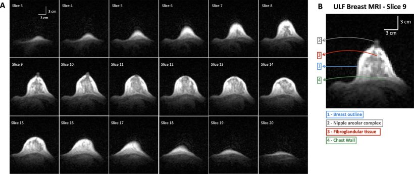
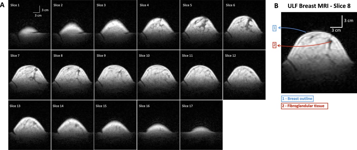
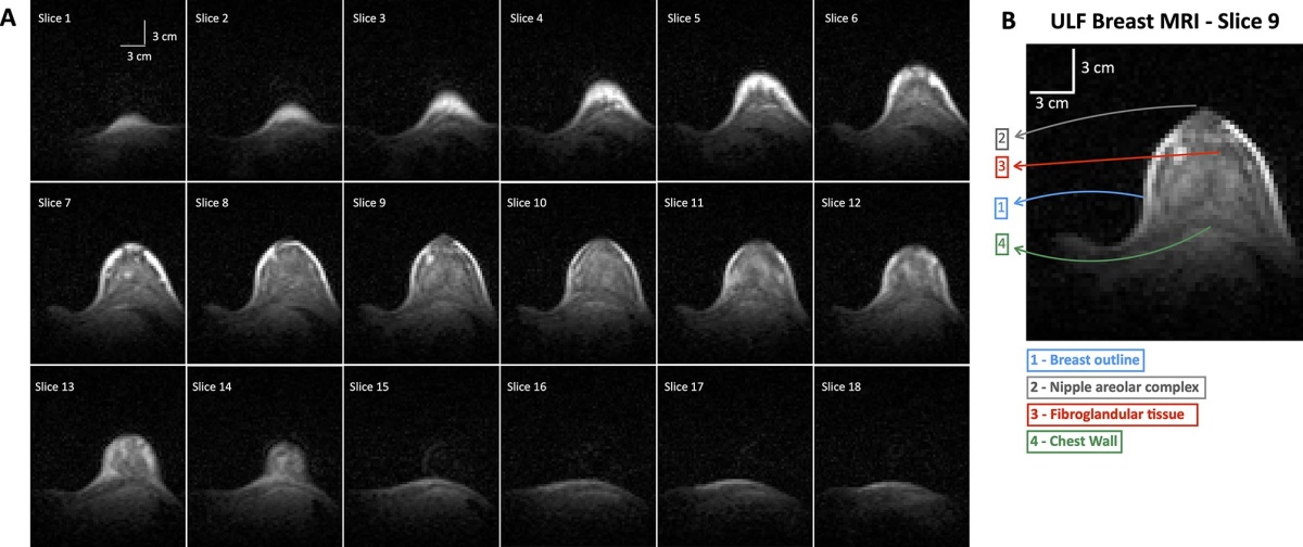
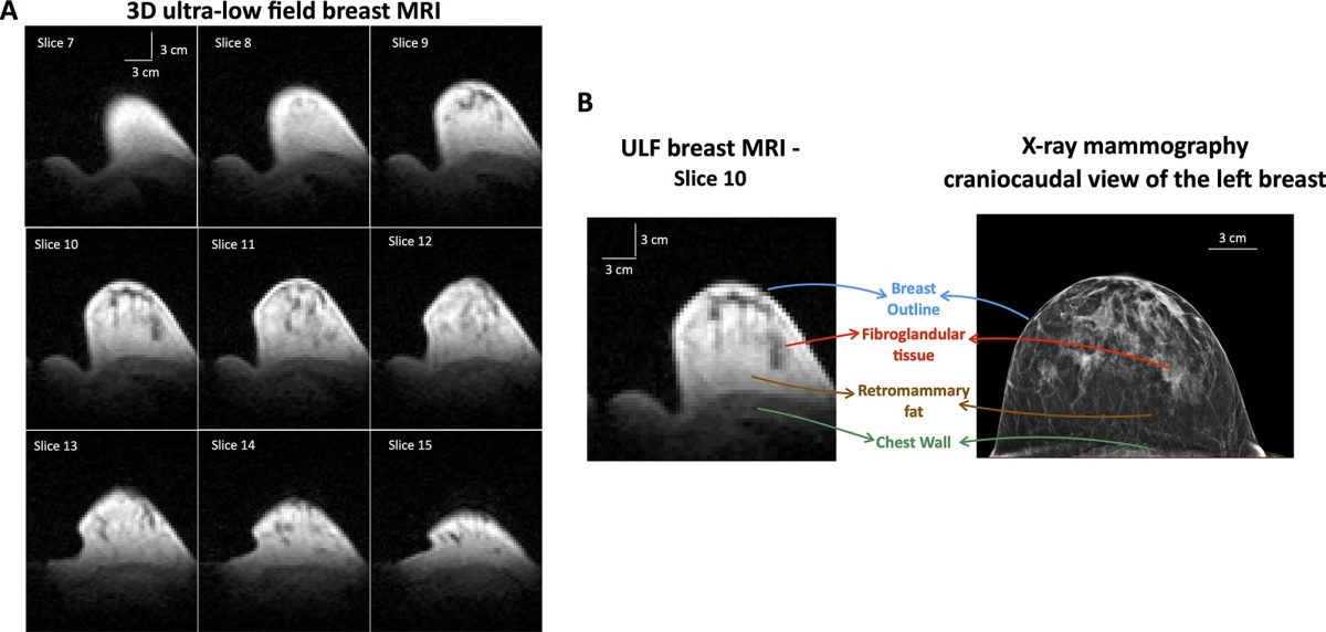
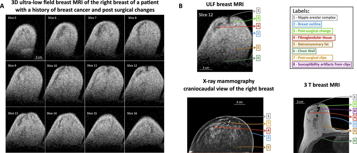
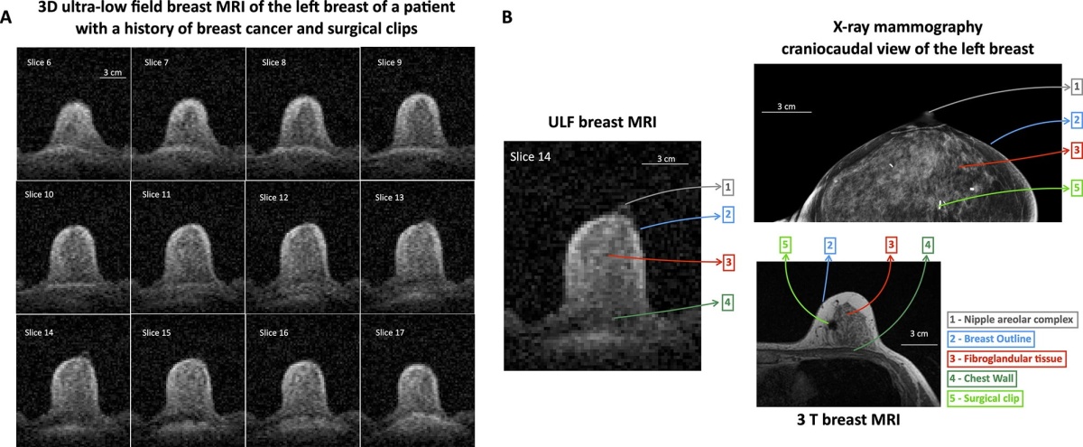
This early evidence suggests that ULF MRI can detect essential breast features and some abnormalities without radiation or injected contrast
Neha Koonjoo
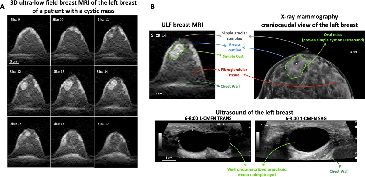
In this study, ULF MRI scans were performed on 14 participants, including 11 women with no history of breast cancer, two women with a prior breast cancer diagnosis, and one woman with a benign mass.
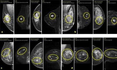
When interpreting the ULF MRI scans, three radiologists could reliably identify essential breast features and distinguish fibroglandular tissue from adipose tissue. The authors note that discrepancies were likely related to the novelty of ULF MRI and may be reduced with additional training and experience.
“This early evidence suggests that ULF MRI can detect essential breast features and some abnormalities without radiation or injected contrast,” said co-first author Neha Koonjoo, PhD, an investigator at the Martinos Center. “These findings point to the potential for ULF MRI as an option that could complement existing screening tools in the future.”
“Even at very low magnetic field, the radiology team was able to make observations about the breast,” said co-principal investigator and co-senior author Kathryn E. Keenan, PhD, from the US National Institute of Standards and Technology. “We attempted this study in hopes that the breast features would be visible, but you don’t always have success. We’re very motivated by this study to continue our work on ultra-low-field MRI for breast screening.”
The researchers note that further study is needed to determine the diagnostic accuracy of ULF MRI for breast cancer screening, including studies in larger cohorts that include patients with benign and malignant lesions. They also emphasize that further refinements in ULF MRI technology are needed to meet clinical resolution standards for breast cancer screening.
“These results will guide the next engineering steps to improve image quality and enable a more comfortable exam and help bring screening to more settings and more patients,” said co-first author Sheng Shen, PhD, of the Martinos Center for Biomedical Imaging.
Source: Mass General Brigham
20.02.2026
