Scientists develop an intelligent knife
Scientists at Imperial College in London, United Kingdom, have developed an ‘intelligent knife’ that instantly informs surgeons whether the tissue they are operating on is cancerous.
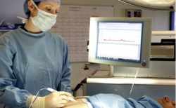
The iKnife is based on electrosurgical knives that use an electrical current to rapidly heat tissue to cut through it. Smoke generated by vaporised tissue is normally sucked away by extraction systems, but iKnife inventor Dr Zoltan Takats realised the smoke was a rich source of biological information. As different types of cell produce thousands of metabolites in different concentrations, he connected an electrosurgical knife to a mass spectrometer and was able to profile the chemicals in a biological sample to reveal information about the state of a specific tissue sample.
In cancers involving solid tumours, surgeons normally take out the tumour with a margin of healthy tissue but acknowledge it is often impossible to tell by sight which tissue is cancerous. For example, one in five breast cancer patients who have surgery need a second operation to fully remove the cancer. With a high level of accuracy reported, the iKnife gives surgeons an immediate answer rather than having to wait up to 30 minutes for laboratory tests results – while the patient is still under general anaesthetic – to know whether tissue is cancerous or not.
‘This is a unique tool because the tissue identification element is built into the cutting device, giving surgeons real-time information about exactly what type of tissue is being cut,’ Dr Takats explained. ‘The intelligent knife will give them an immediate answer, enabling them to continue the operation without interruption.’
Studies showed the iKnife was 100% accurate when diagnosing tissue samples from 91 patients and matching the post-operative diagnosis based on traditional methods.
Researchers have also used the iKnife to analyse tissue samples collected from 302 surgery patients, recording the characteristics of thousands of cancerous and non-cancerous tissues - including brain, lung, breast, stomach, colon and liver tumours - to create a reference library. The iKnife works by matching its readings during surgery to the reference library to determine what type of tissue is being cut, producing a result in less than three seconds.
Dr Takats, an analytical chemist at ICL, said: ‘These results provide compelling evidence that the iKnife can be applied in a wide range of cancer surgery procedures. It provides a result almost instantly, allowing surgeons to carry out procedures with a level of accuracy not possible before. We believe it has the potential to reduce tumour recurrence rates and enable more patients to survive.’
He added that the benefits to patients include improved accuracy, meaning that resection is kept to a minimum, as well as reduced exposure to anaesthetic.
The iKnife is not commercially available at this stage. Whilst its accuracy has been proven in trials, Dr Takats said the next step is for a clinical trial to see whether giving surgeons access to the iKnife can improve patient outcomes.
Although the current study focused on cancer diagnosis, Dr Takats says the iKnife can identify other features, such as tissue with an inadequate blood supply or types of bacteria present in the tissue.
The National Institute for Health Research (NIHR) Imperial Biomedical Research Centre, the European Research Council and the Hungarian National Office for Research and Technology funded the study.
Lord Darzi, Professor of Surgery at ICL and the study’s co-author, said: ‘In cancer surgery, you want to take out as little healthy tissue as possible, but you have to ensure that you remove all the cancer. There is a real need for technology that can help the surgeon determine which tissue to cut out and which to leave in. This study shows that the iKnife has the potential to do this. The impact on cancer surgery could be enormous.’
Profile:
A former post-doctoral research associate at Purdue University in Indiana, USA, as well as Director of the Cell Screen Research Centre and Head of Newborn Screening and Metabolic Diagnostic Laboratory at Semmelweis University, Budapest, Dr Zoltan Takats later became a Junior Research Group Leader at Justus Liebig University, Giessen, Germany. In 2012 he moved to the United Kingdom where he is a Reader at Imperial College, London. He has pioneered mass spectrometry research and is one of the founders of Ambient Mass Spectrometry, as well as primary inventor of six mass spectrometric ionisation techniques and the founder of several companies that pursue analytical and medical device development..
05.11.2013