Rise of the machine: Ultrasound technology crushing professional development and research
The past year has not improved the prospects for ultrasound, according to Lorenzo Derchi, MD, who chaired the session at ECR 2010 dedicated to « Future direction of ultrasound » and opened the discussion by saying the field is in decline.
« There are further advances with contrast agents, volume scans and image enhancement techniques, » he said.
« Yet as a profession, the perspectives for ultrasound only seem worse, » he said, noting that the number of presentations given at the Radiology Society of Noth America (RSNA) and here at ECR have declined again this year.
Stopping short of saying there is a crisis, « Ultrasound faces professional development and research problems, » said Dr. Derchi, who is a past president of the European Society of Urogenital Radiology,
In 2009 Derchi raised eybrows, and some objections, with an article entitled « Ultrasound: a strategic issue for radiology? » published in the journal of the European Society of Radiology.
At Saturday's session he repeated his provocation that « the relationship between ultrasound and radiology has not been an easy love story, » and went on to decry the continual focus on ultrasound techiques to the detriment of the development of ultrasound as a medical profession.
« As a result, the routine services of ultrasound are guaranteed, but for young doctors it is only seen as a mature technique with no room for advancement, » he said.
Ultrasound is increasingly introduced to clinical practice by non-radiologists and as for its future direction, it seems doomed to be regarded as only a stethoscope for the majority of practitioners.
Michel Claudon, who co-authored the controversial article with Dr. Derchi in 2009, again supported his colleague agreeing with his conclusions.
Dr. Claudon with the Radiology group at the University of Nancy in France, then indirectly demonstrated the fast development of this technique by asking for a show of hands from the audience for those currently using contrast agents in examinations.
To his surprise two-thirds of the participants have used this novel approach for which guidleines were published in 2008.
After a review of studies and cases for applications of ultrasound contrast in liver and kidney, Dr. Claudon concluded that this technique's principal application is as a complement to contrast-enhanced computed tomography (CE-CT) or magnetic resonance imaging (CE-MRI) in pretreatment and that it is emerging as a major tool for follow on studies for vaidation of therapies and especially for recurrence in cancer patients.
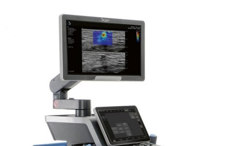
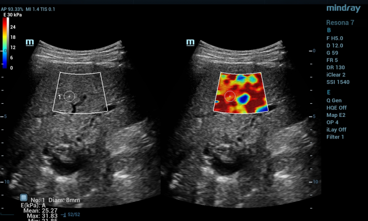
Moving into yet another emerging technology in ultrasound, Giorgio Rizzatto, MD, with the University of Udine, took on the question « Elastography: Clinical tool or toy? »
On the way to giving his answer, Dr. Rizzatto sounded a grim note for the compression technique that pioneered the advances of elastography for measuring tissue stiffness, saying « compression is over, » with the advance of new technologies that produce a shear wave to reliably and reproducibly quantify the extent of tissue stifness.
Noting that current evidence is crushed by the initial focus on breast studies, elastography holds promise for a braod range of clinical applications for eyes, skin, muscles, liver prostate, vessels and the heart.
Dr. Rizzatto concluded that elsatography will prove to be a clinical tool, and not a toy, for routine practice, screenings and monitoring patient conditions.
But he emphasized that elastography is « only a decriptor and not the Bible, » and is considered as a complement to other modalties.
Simon Elliot, MD, Radiologist, Clinical Lead in Ultrasound at the Freeman Hospital, Newcastle upon Tyne described the diagnostic advantages emerging in his presentation « Volume US in clinical practice for the radiologist. »
Pointedly, he said, his focus was on non-obstetric work as one of the first applications for capturing three dimensional volumetric data sets was to render «pretty baby faces in the womb. »
« Actually, we do not often look at these 3D reconstruction images in the clinic, » he said. « Granted they are a «'Wow' feature of the software, but there is little diagnostic value beyond providing a perception of the target organ. »
Instead, he said, the focus is upon the three different 2D plane views rendered by the data sets that can then be viewed at any rference point on the x, y and z axes.
Dr. Elliot suggested that in practice his group is using volume acquisition to speed up routine exams that formerly were done with the conevntional 2D mode.
«
« In 90 seconds scan time we have a massive block of data and can take measurements or scroll through different views, » he said.
06.03.2010


