News • iPS cell-derived mesenchymal stem cells
Researchers discover new way to generate high-quality cartilage
A Japanese research group, led by Dr. Denise Zujur and Associate Professor Makoto Ikeya, has developed a method using iPS cell-derived mesenchymal stem cells (iMSC) to create cartilage spheroids, offering new possibilities for tissue repair.
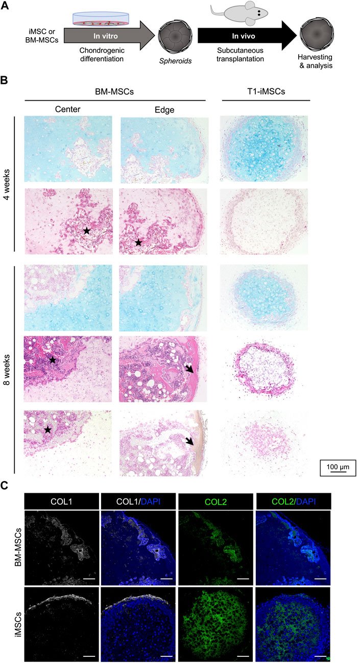
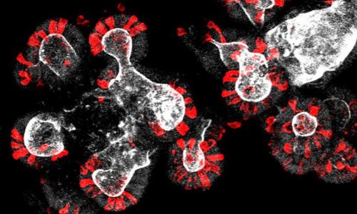
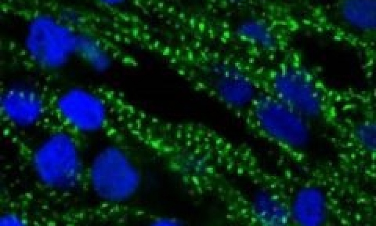
Image source: Zujur et al., Frontiers in Cell and Developmental Biology 2023 (CC BY 4.0)
Current treatments for cartilage tissue repair are limited, and regenerative medicine commonly utilizes cartilage and mesenchymal stem cells. However, clinical applications of these cell types are limited by medical complications from donors and poor proliferative capacity.
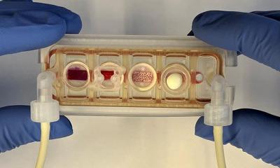
To overcome these challenges, the research team explored a differentiation method to generate high-quality cartilage spheroids from iMSCs. They discovered that using a small molecule compound called TD-198946 improved the efficiency of differentiating iMSCs into cartilage. By employing a stepwise induction protocol, they successfully produced high-quality cartilage spheroids. They further demonstrated that these spheroids, when transplanted into immunodeficient mice, increased extracellular matrix production, indicating the maintenance of cartilage in vivo. Furthermore, the research team found no signs of dedifferentiation, fibrotic cartilage formation, or hypertrophy in vivo, thus verifying the safety for potential clinical use.
The results of this study were published online in Frontiers in Cell and Developmental Biology.
This breakthrough highlights iMSCs as a promising cellular source for cartilage repair using stem cells. Additionally, the spheroids' ability to fuse within a few days opens the possibility of using bioprinting techniques to construct larger cartilage tissues. This research offers new avenues for cartilage repair and provides a novel means to generate significant quantities required for therapeutic cartilage transplantation. Future research could explore the feasibility of clinical applications and investigate the potential of combining bioprinting technologies with iMSC-derived cartilage spheroids for large-scale tissue reconstruction.
Source: Center for iPS Cell Research and Application, Kyoto University
19.06.2023