Real-time Tissue Elastography
A bridge between mammography and biopsy
Radiologists are paying increasing attention to ultrasound real time tissue elastography (HI-RTE). Earlier this year, at their annual meeting, the Austrian, Swiss and German Ultrasound Societies (ÖGUM, SGUM, DEGUM), highlighted the effectiveness of the method in differentiating soft from stiff tissue, i.e. healthy areas from tumours, a key differentiation in breast cancer diagnosis.

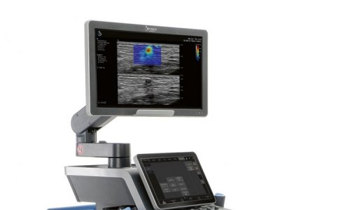
Meeting with Ellison Bibby, European Product Manager for Radiology Ultrasound with Hitachi Medical Systems, Daniela Zimmermann, of European Hospital, asked for an update on the use of sonoelastography in this area.
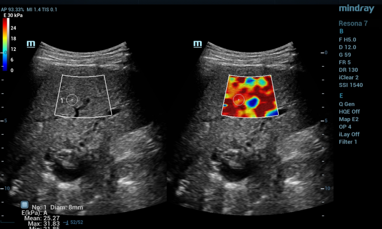
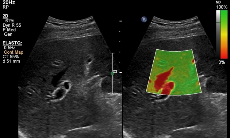
The breast is the best-known example of how patients are taught to palpate for tumours. A tumour is stiff compared to the surrounding tissue, and that’s exactly what real-time tissue elastography images,’ Ellison Bibby explained. ‘The stiffness relative to the surrounding tissue. It is similar in the prostate gland: the digital rectal examination is used to palpate the prostate for hard lumps and, on the elastography image, we can see areas that are stiffer. Normal ultrasound gives us information about the anatomy and reflectivity of the tissues, but HI-RTE is like a ‘palpation image’, that looks at new additional information – how the tissues react to stress, which is related to tissue elasticity’.
The tissues are gently compressed by the transducer in a repetitive manner to distort the tissue. ‘The echo data from one image frame is compared to the next to build up a pattern of tissue displacement. Where there are differences in tissue stiffness, the amount of displacement varies – normal soft tissues will show greater distortion than stiffer tissues’.
Asked about the field of work in which this technique can be used, Ellison Bibby explained that if, after mammography and an ultrasound examination, the results are still uncertain, elastography can be used to provide further differentiation. ‘It can build a bridge between mammography screening programmes and additional MR- examinations or biopsies. Today, this is a very sensitive area and very few doctors would be convinced to dispense with the biopsy, even when the real-time tissue elastography results look perfectly benign. But, several published studies, with quite large numbers of patients have shown comparable results, which, in those patients that are of low suspicion for cancer, elastography could be used to decide which strategy to follow: biopsy or follow-up imaging after a short interval. But, of course we need more studies before we reach the stage of replacing biopsy with sonoelastography’.
Today, the BI-RADS categories, based on imaging appearances, are used for screening, but there are problems in terms of reproducibility, especially for BI-RADS 3 (probably benign) and BI-RADS 4a (low suspicion of cancer). It has been reported that approximately 70% of patients with benign lesions undergo further investigation – either short term follow up, aspiration or biopsy. Our goal is to show convincingly in this group of patients, that when we have a benign result with sonoelastography we could effectively downgrade the BI-RADS score by 1, giving sonoelastography a definitive role in breast cancer diagnosis.’
20.12.2008