Image source: IBA Dosimetry
Article • From geometric phantoms to clinically predictive testing
The evolving role of phantom-based QA in medical imaging
Quality assurance (QA) in medical imaging has traditionally focused on the technical performance of scanners, ensuring consistent output within defined limits. Conventional QA methods typically use simple test objects made of uniform materials and basic geometries.
These geometric phantoms offer reproducible benchmarks independent of patient variability. However, their very simplicity is also their limitation, as they do not reflect the anatomical complexity encountered in clinical practice. As imaging technology evolves and artificial intelligence (AI) becomes integrated into routine workflows, the gap between conventional QA and real-world clinical performance has grown increasingly apparent.
Modern imaging systems employ advanced acquisition and post-processing techniques that interact with anatomical structures and affect both image quality and diagnostic interpretation. In this setting, traditional metrics derived from geometric phantoms often fall short of predicting actual clinical performance. QA is therefore shifting from purely technical measurements to outcome-oriented frameworks that assess whether diagnostic objectives – such as accurate lesion detection – are consistently met. The growing use of AI in imaging further underscores this need. AI algorithms are sensitive to variations in image acquisition and frequently operate as opaque black boxes, making controlled and clinically relevant validation essential. Phantom-based testing is uniquely positioned to address these challenges, provided that phantoms capture not only the physics of imaging but also the anatomical and pathological features relevant to diagnostic tasks.
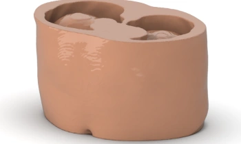
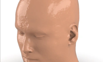
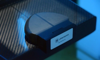
In response to these evolving requirements, a new class of anthropomorphic phantoms has been developed. PhantomX is among the organizations advancing this approach, producing phantoms designed to replicate human anatomy, tissue heterogeneity, and disease characteristics relevant to diagnostic imaging. Enabled by advances in materials science and manufacturing, these phantoms extend beyond the uniform structures of traditional designs by incorporating features that mimic bone, soft tissue, organ structures, and pathologies (Figure 1). This increased realism allows imaging systems and AI-based analysis workflows to be evaluated under conditions that closely reflect clinical practice, bridging the gap between physical quality assurance and clinical relevance (Figure 2).

Image adapted from: Goelz et al. (2025), Scientific Reports (CC BY 4.0)
A major advantage of using physical anthropomorphic phantoms in this context is reproducibility. While clinical data can be variable and influenced by patient-specific factors, a physical phantom can be imaged repeatedly under controlled conditions. This enables standardized comparisons across devices, sites, and time points. It also supports traceability – a growing requirement in the evaluation of AI tools, where regulators increasingly expect transparent evidence demonstrating how an algorithm performs under different scenarios. As a stable reference for proactive on-site validation and surveillance, physical phantoms can complement population-based AI evaluation and strengthen the robustness and interpretability of performance assessments.
The development of realistic anthropomorphic phantoms has also opened pathways for new kinds of research collaborations. PhantomX has partnered with clinical and scientific groups, including consortia focused on breast imaging, algorithmic performance, and institutions responsible for large-scale CT quality management. Such collaborations have leveraged the phantoms' ability to replicate clinically relevant structures for testing, education, and AI deployment. These partnerships demonstrate how standardized physical models can facilitate multi-center studies, support method comparison, and ensure that rigorous algorithmic surveillance translates into robust performance.
In recent years, the importance of this approach has been further recognized in industry and regulatory discussions. As recent as November 2025, PhantomX has partnered with IBA Dosimetry GmbH, a provider of dosimetric and quality assurance solutions for medical imaging and radiation therapy. The integration reflects a broader commitment to advancing QA technologies capable of supporting both traditional imaging and emerging AI-driven workflows. There is a clear need for tools that improve safety, effectiveness, and transparency in medical imaging – a vision strongly aligned with the evolving expectations of clinical physicists and regulatory agencies.
What makes PhantomX phantoms particularly valuable for the medical physics community is that they can serve as a common testing platform, independent of vendor ecosystems, scanner generation, or software version. As AI becomes more deeply embedded into acquisition, reconstruction, and diagnostic pathways, the need for such neutral, standardized testing environments will continue to grow. Medical physicists are increasingly tasked with evaluating not only hardware performance, but also how intelligent systems contribute to – or potentially bias – clinical decision-making. Here, realistic anthropomorphic phantoms offer a stable foundation for generating the kind of evidence that supports safe adoption.
In summary, the introduction of anatomically realistic physical phantoms reflects a meaningful shift in imaging QA from a primary focus on technical scanner performance toward more outcome-oriented evaluation frameworks. By enabling the evaluation of imaging systems under controlled yet clinically realistic conditions, these phantoms bridge the gap between conventional performance metrics and the lived clinical reality in which both humans and AI operate. As radiology continues to evolve into a more complex and data-driven discipline, such tools will play an increasingly important role in ensuring safe, effective, and transparent imaging practices.
The authors

Arianna Giuliacci, Nuclear Engineer, head of the Clinical Application team at IBA Dosimetry. More than 15 years of experience as R&D physicist, customer manager, providing clinical implementation of IBA products.

Dr. Paul Jahnke, Radiologist and founder of PhantomX
Source: IBA Dosimetry
(First published in EFOMP Magazine)
12.09.2026