New ways to treat slipped discs
St. Joseph Hospital in Essen, Germany, has brought hope to patients with previously inoperable or untreatable slipped disks, either because they suffered arthritis, or conventional surgery meant high-risk.
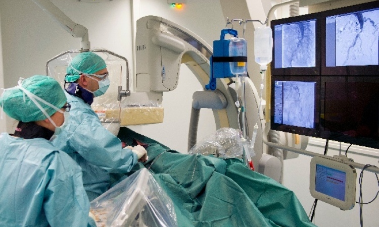
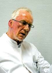
For the last nine months, Jens Timmermann MD PhD, Head of the Interventional Radiology Department, has been using micro-invasive technology to treat these patients.
up a multitude of
new opportunities
This enables him to access the intervertebral discs despite severe osteoarthritis, while simultaneously taking a tissue sample to determine the degree of disc degeneration and customise treatment.
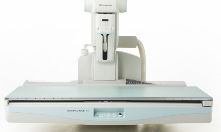
Apart from the surgeon’s skill, today X-ray technologies in particular facilitate such innovative and minimally invasive procedures. At the hospital such interventions as well as diagnostic examinations are carried out in the specialist radiological surgery, named diavero, which is linked to the hospital. Frank Stöblen MD, Specialist for Diagnostic Radiology, and Dr Karlgeorg Krüger, both heads of diavero, are among the first in Europe to use the latest angiography equipment, made by the Japanese firm Shimadzu, in the field of intervention, and particularly for Dr Timmermann’s slipped disc treatment.
We visited these medical pioneers in the Ruhr Area to discuss the advantages of the procedure and developments that could further improve slipped disc treatments.
EH: Interventional radiology is already using laser technology for the treatment of slipped discs. What is different about the micro-invasive procedure which you are employing and what are the advantages?
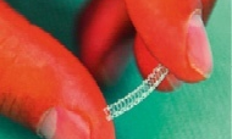
Jens Timmermann (JT): A chamber system was developed in joint cooperation with an American company. This contains a cannula and is navigated into position with the help of a spiral catheter. This cannula allows us access to the disc. We produce low pressure in the chamber and at the same time inject a saline solution. The disc material, which is sucked in, is cut and washed out of the cannula. The advantage, compared to the laser procedure, is that there is no burned tissue next to the spinal cord and the tissue can be differentiated and examined in pathology. This means we can cover a very broad spectrum of patients and also treat patients whose only other alternative would have been surgical stiffening of the vertebral body. The procedure makes it possible to operate on slipped discs in the neck area with micro technology, lowering the usual rate of complication associated with open surgery (20-30%) to a mere minimum. Therefore, we can treat people who would otherwise face a life with significant limitations. There are very few places in Europe where this procedure is carried out. However, in America and Asia it is much more common. This is, among other reasons, due to the high interest in, and standard of, technology in Asia.
What are the technological prerequisites for this intervention?
Frank Stöblen: You need real high-end equipment with excellent image quality – with low radiation. The interventions carried out by Dr Timmermann take, on average, 45 minutes. Even if the work is carried out using radiology images, and the patient is not exposed to the full radiation dose for the entire time, this factor still plays a significant role. In Essen we work with the latest generation of Shimadzu angiography systems and are very happy with them, particularly in terms of the radiation dose. Inspection by the Technical Inspection Authority showed that the system produces only a third of the dose produced by comparable systems, surely a result directly linked to the direct-conversion flat panel detector, a particular feature of the Shimadzu equipment.
A further advantage is reliability, which we had already experienced with the predecessor. The system has no down-times and is continually used without any technical failures. Another decisive feature is a programme that very quickly produces a multidimensional image of the catheter tip. This is otherwise very hard to visualise; although some angio-CTs have this function it takes several minutes – compared to just a few seconds with our angiography system.
Which other therapeutic perspectives does the micro-invasive procedure offer?
JT: The next step, which we are already in the process of taking, is that, on the basis of the tissue sample removed, we grow new tissue and re-implant it. The application for this procedure was approved by the Medical Association last year and we have found a biogenetic laboratory in Berlin that can grow the tissue. We have now successfully carried out the re-implantation in four patients. And next — the new micro-invasive procedures open up a multitude of new opportunities from which we’ll benefit a lot over the next few years.
01.03.2009