News • In heart disease

Lab-grown pig heart tissue could replace animals research

A new way to replicate what happens inside the heart after cardiac arrest could open new avenues for the study of heart regeneration whilst reducing the use of live animals in research, according to a study from the University of Surrey and King’s College London.

Photo
Fig. 1: Porcine epicardial slices preparation. Schematic representation of porcine ventricles sampling and slicing. Heart collection and setup: (1) Heart is harvested maintaining the pericardium and retroperfused with ice-cold cardioplegia solution. Ventricles are then separated from the heart avoiding touching the epicardium, to prevent damage. (2) Embedding area must be set up prior to tissue slicing to allow all components to cool down to ice-cold temperature. Tissue block preparation: (3) Cardiac tissue is placed on the ice-cold Petri dish and (4) blocks of tissue 8 mm ×8 mm are dissected with a single edge steel blade. Tissue block embedding: (5) The epicardial side of the slice is flattened onto a cold agarose surface and placed in the middle of a custom-made 3D printed ring. 5–8 ml of 30–37 °C low melting agarose is poured to embed the tissue block. (6) Once solidified, the block is removed from the ring and (7) squared on one side to allow a better alignment with the vibratome’s blade (red dashed line). Slice cutting and culture: (8) Embedded tissue blocks are oriented along the squared side and glued to the vibratome specimen holder. The blade is aligned to the epicardial surface and 400–500 µm thick slices are cut using a high precision vibratome. (9) Slices are allowed to recover for at least 30 min in a recovery bath. (10) Epicardial slices are cultured epicardium-up on 8-mm-high pillars cast at the bottom of a Petri dish. (11) En face immunohistochemistry of WT1 (red) and mesothelin (MSLN, green) shows the presence of a continuous epicardial layer, characterized by typical morphology and marker expression (WT1+ cells indicated with full arrowhead, WT1− MSLN+ cells indicated with empty arrowhead). This figure was created using BioRender.com.
Source: npj Regenerative Medicine (2022). DOI: 10.1038/s41536-021-00202-7

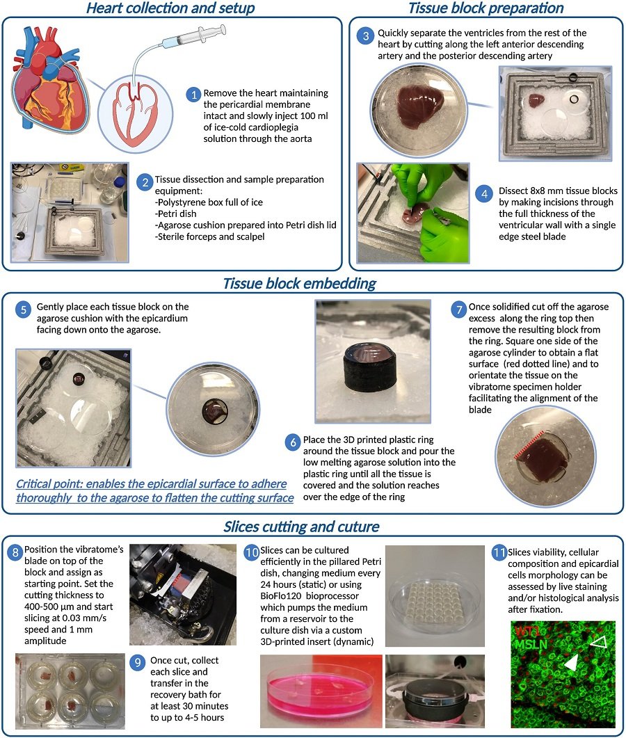
Researchers at the University of Surrey and King’s College London have developed a process that involves obtaining and growing thin slices of pig heart tissue which include both the epicardium - the most external layer of the heart that contains cells that can promote its recovery - and underlying heart muscle.

The team treated the epicardial slices with stimulating compounds, showing that cells become activated in a way that replicates what happens in the heart after a heart attack. The new process was able to reproduce observations typically obtained in live animal models.

Dr Paola Campagnolo, lead author of the study and Senior Lecturer in Molecular Cardiovascular Sciences at the University of Surrey, said: “This research typifies the One Health, One Medicine ethos at the University of Surrey, as our model could help us understand how to stimulate the repair process after heart attacks without the need to use live animals in the research. We are hopeful our model could lead to better health outcomes for humans and reduce the reliance on animal experiments in cardiovascular science.”

According to the British Heart Foundation, there are around 7.6 million people living with heart or circulatory disease in the UK. This disease causes a quarter of all deaths in the UK.

The ability of the heart to recover after an injury is severely limited by the low number of regenerating cells within its tissue. The current research models and strategies aimed at improving the heart’s repair process are mainly based on surgical procedures performed on laboratory animals.  

Dr Davide Maselli, Postdoctoral Research Associate and first author of the paper, said: “This work provides an innovative tool to study the healing from a heart attack. We believe that our model could be useful to dissect the role of different cells in the reparative process. In our consideration, it is extremely important that every step forward in this field delivers a clinical perspective for the patients while reducing the burden on research animals.”

The research, published in the journal npjRegenerative Medicine, proposes a system to study the regeneration of the heart in a laboratory dish and could therefore lead to a reduction in the number of small animals used in cardiovascular research.

Source: University of Surrey

13.03.2022

Read all latest stories

Related articles

Photo

News • Ventricular fibrillation research

Promising therapeutic target for cardiac arrhythmias discovered

A newly discovered signaling mechanism implicated in the development of ventricular fibrillation offers promise of future treatment options for this life threatening condition.

Photo

News • Heart perfusion simulator

iHEART: a new model to boost cardiac research

A team at Politecnico di Milano developed iHEART, a mathematical and computational model of the human heart designed for studying coronary artery disease.

Photo

News • Economic burden of CVD

Putting a price tag on cardiovascular disease in Europe

Cardiovascular disease (CVD) cost the EU an estimated €282 billion in 2021, according to late breaking research presented at the European Society of Cardiology's ESC Congress 2023.

Related products

Subscribe to Newsletter