News • Light it up
Faster, more accurate cancer detection using nanoparticles
Light-emitting nanoprobes can detect cancer early and track the spread of tiny tumors
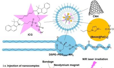
Using light-emitting nanoparticles, Rutgers University-New Brunswick scientists have invented a highly effective method to detect tiny tumors and track their spread, potentially leading to earlier cancer detection and more precise treatment. The technology could improve patient cure rates and survival times. “We’ve always had this dream that we can track the progression of cancer in real time, and that’s what we’ve done here,” said Prabhas V. Moghe, a corresponding author of the study and distinguished professor of biomedical engineering and chemical and biochemical engineering at Rutgers–New Brunswick. “We’ve tracked the disease in its very incipient stages.”
The Achilles’ heel of surgical management for cancer is the presence of micro metastases
Steven K. Libutti
The study, published online in Nature Biomedical Engineering, shows that the new method is better than magnetic resonance imaging (MRI) and other cancer surveillance technologies. The research team included Rutgers’ flagship research institution (Rutgers University-New Brunswick) and its academic health center (Rutgers Biomedical and Health Sciences, or RBHS). “The Achilles’ heel of surgical management for cancer is the presence of micro metastases. This is also a problem for proper staging or treatment planning. The nanoprobes described in this paper will go a long way to solving these problems,” said Dr. Steven K. Libutti, director of Rutgers Cancer Institute of New Jersey. He is senior vice president of oncology services for RWJBarnabas Health and vice chancellor for cancer programs for Rutgers Biomedical and Health Sciences.
The ability to spot early tumors that are starting to spread remains a major challenge in cancer diagnosis and treatment, as most imaging methods fail to detect small cancerous lesions. But the Rutgers study shows that tiny tumors in mice can be detected with the injection of nanoprobes, which are microscopic optical devices, that emit short-wave infrared light as they travel through the bloodstream – even tracking tiny tumors in multiple organs. The nanoprobes were significantly faster than MRIs at detecting the minute spread of tiny lesions and tumors in the adrenal glands and bones in mice. That would likely translate to detection months earlier in people, potentially resulting in saved lives, said Vidya Ganapathy, a corresponding author and assistant research professor in the Department of Biomedical Engineering.

“Cancer cells can lodge in different niches in the body, and the probe follows the spreading cells wherever they go,” she said. “You can treat the tumors intelligently because now you know the address of the cancer.” The technology could be used to detect and track the 100-plus types of cancer, and could be available within five years, Moghe said. Real-time surveillance of lesions in multiple organs should lead to more accurate pre- and post-therapy monitoring of cancer. “You can potentially determine the stage of the cancer and then figure out what’s the right approach for a particular patient,” he said.
In the future, nanoprobes could be used in any surgeries to mark tissues that surgeons want to remove, the researchers said. The probes could also be used to track the effectiveness of immunotherapy, which includes stimulating the immune system to fight cancer cells.
Source: Rutgers University-New Brunswick
13.12.2017


