Breathing space
If the hopes of inventors are to be believed, in around 20 years’ time there will be ‘real artificial lungs -- for now the endpoint of a history that began 84 years ago with the invention of the iron lung. Until then, non-invasive and invasive mechanical respiration will continue to dominate the hospital, complemented by extracorporeal procedures for blood oxygenation and decarbonisation, writes Holger Zorn.
In the autumn of 1928 the starter’s gun fell for the first clinical lung replacement procedure: a hollow tube made from metal into which the patient was positioned, closed airtight at the neck and abdomen. Rhythmic changes of air pressure in the tube led to chest expansion and constriction and air entered the lungs in the normal way via the mouth. Although it was not possible to treat diseased lung tissue with the help of the ‘iron lung’, it was possible to treat paralysed respiratory muscles – and plenty of patients needed this treatment. Just into the century a known virus began to change – poliomyelitis.
Known previously as a disease that occurred only sporadically, it now swept across continents like a pandemic. With the help of the iron lung it was possible to support the breathing in children who suffered acute symptoms of paralysis until their respiratory muscles recovered. However, some patients never recovered; they depended on this technology for the rest of their lives. The iron lung therefore demonstrated the consequences of maximum intensive therapy and is still synonymous with being a cold, steely prison. Exactly 60 years ago around 30 patients a day were being admitted to the Copenhagen Municipal Hospital with respiratory paralysis during a new outbreak of the disease. In view of this pressure, anaesthetist Björn Ibsen developed an alternative treatment whereby patients were intubated and ventilated over long periods of time with bags. Two years later, Ibsen also founded the world’s first intensive care ward in the same hospital.
Invasive ventilation
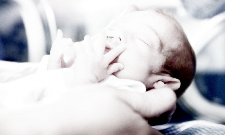
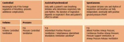
These days, modern ventilators with special tube systems facilitate lung ventilation for premature babies without damaging the yet immature tissue – lungs of babies where the volume corresponds with the size of a plum. Computerisation in medical technology, together with an increasingly better understanding of lung physiology, has resulted in continuous innovations. Ventilation modes designed to monitor the volume were combined with pressure-controlled modes. There are now special ventilation procedures for all the different diseases of the lung (see table).
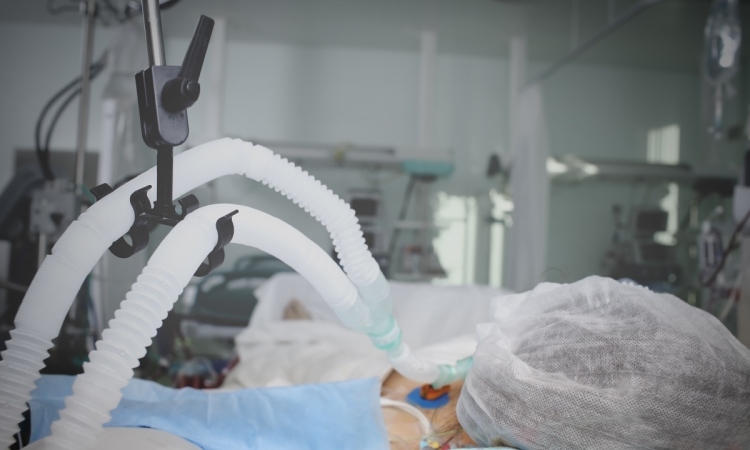
Current machines are capable of specialist manoeuvres that open closed areas of the lung. With this so-called open lung technology the ventilator temporarily increases pressure in the lungs to a level for only a short time, which, if maintained for a long time, would cause great damage. Closed areas in the lungs then open and can be ventilated. The machine fastidiously ensures that there is never a neutral or even negative pressure at the end of the following ventilation cycle. The positive end-expiratory pressure protects these areas from renewed collapse. These days, the computer also takes over large parts of the process involved in weaning patients off the equipment. It recognises any attempt the patient makes to breathe on his own and even supports this. The weak respiratory muscles begin with the intake of breath, but if this intake is too flat the ventilator completes the process – always with a guarantee that, if the patient is completely exhausted, the machine will take over again completely.
Non-invasive ventilation
Another approach is the use of airtight facemasks. Using the most up-to-date algorithms they ensure that patients who are awake are supported in all their breathing. Small leaks are detected and compensated for. Such non-invasive types of ventilation can, for instance, support patients with coronary disease who suffer from fluid on the lungs. The risks of a long weaning process from ventilation or of an anaesthetic, which would be an additional strain on the patient’s cardiovascular system, are thus avoided.
Extracorporeal ‘ventilation’
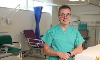
If the lung tissue itself is incapable of working at all then even the most modern ventilator cannot help, says Michael Rühl of the University Hospital Greifswald. ‘We use special membranes here, which imitate lung function. As with the pulmonary alveoli, the gasses here permeate from one side of the membrane to the other. Blood low in oxygen is directed out of the body through tubes and flows past this multi-layered membrane, which, despite its small dimensions, has the surface area of a football pitch. It is then oxygenated and the carbon dioxide is removed before it flows back into the body.’
Mechanical ventilation
Clotting has always been a problem with this process; however, surfaces now facilitate oxygenator use over several days. Lung injuries or viral diseases are the most typical indications for this treatment in adults, along with infections amongst intensive care patients. The German ECMO Centres always have a few more patients during the annual flu season, but this treatment is very invasive, expensive and not free of side effects: ‘An ECMO,’ says Michael Rühl, ‘is only connected if there is the chance of a cure, or if lung transplantation can be carried out.’ Many new procedures are being trialled, and not all make it to the clinical application stage. The so-called ‘liquid ventilation’, originated in deep sea diving, was meant to treat respiratory failure from around the turn of the millennium. This procedure involves perfluorocarbons (fluids which, strongly enriched with oxygen, are to achieve better lung oxygenation) being nebulised in the breathing gas. Although some studies have shown the effectiveness of the method, it is not superior to modern ventilators. In fact, none of the procedures are an adequate substitute for a real lung.
Artificial lung
At least the issue of material required for a possible artificial lung has now been resolved. Robert A Potkay of the Advanced Platform Technology Centre, Louis Stokes Cleveland VA Medical Centre, has developed a silicone rubber that contains extremely fine canals (see image), similarly to those in the lungs. The possible diffusion distance is very small whilst the surfacevolume ratio is high. This means that the lung could be supplied with simple air rather than oxygen, and this with a size that would not hamper an implantation (source: Lab Chip; DOI 10.1039/c1lc20020h). The development of this tissue is the first step because, whilst the heart could pump blood into the new lung, the chest muscles would have to be able to expand the tissue via negative pressure in order to replace the original – an enormous challenge for the support apparatus.
Therefore, the replacement of one of the most complex human organ systems has not yet been achieved.
02.05.2012