A multimodal approach strengthens non-invasive diagnostics
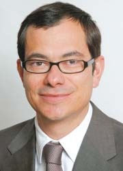
Matthias Gutberlet, Professor of Cardiac Imaging at Leipzig University and head of the Department for Diagnostic and Interventional Radiology at the Heart Centre in Leipzig, welcomed radiologists, cardiologists and nuclear medicine experts, in March, to the 2nd Non-invasive Cardiovascular Imaging Symposium. Their primary focus was on technological opportunities in cardiac and vessels imaging. Non-invasive procedures such as echocardiography, MRI, CT or myocardial scintigraphy already cover a broad spectrum in cardiovascular diagnostics and these will grow; a trend away from purely diagnostic interventions is clear. In an interview with Meike Lerner (European Hospital), Prof. Gutberlet summarised the most significant advances in imaging modalities, as well as their potential and limitations
Echocardiography is the work horse of non-invasive cardiovascular diagnostics. Has this developed?
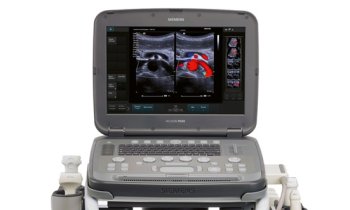
Matthias Gutberlet: 3-D-echocardiography in real time has proved a useful tool, particularly in cardiac surgery to assess heart valves before and after reconstruction. 3-D-echocardiography has also shown promising potential for the imaging of the left ventricle. However, ultrasound procedures have their technological limitations, which is why we frequently dependent on other imaging procedures because of insufficient sonographic quality, particularly in the imaging of the right ventricle. To counteract existing weaknesses of individual non-invasive procedures, there is definitely a trend towards multimodal strategies.
Which procedures are included in those multimodal strategies?
This depends largely on the individual indication: MRI certainly delivers the most information, such as on tissue differentiation, vitality, function etc. However, there are also attempts at recording 3-D data sets through higher field strengths and faster imaging, to allow us to carry out reconstructions similar to those done using CT.
CT is currently particularly suitable for coronary diagnostics but, compared with the invasive heart catheter, is currently used relatively infrequently. I’m sure this will change in the future because the development of equipment with a lot more than 64 slices and improved rotation times will significantly increase the time resolution. Whereas achieving an image of the entire heart with the 64-slice CT normally still lasts over several heart beats the latest generation equipment can partly reduce this time down to just one heart beat. This has enormous advantages, both for the diagnosis of patients with higher heart frequency as well as regarding radiation exposure. Combined with perfusion analysis, i.e. an ischaemic indication for the clarification of haemodynamic relevance for the narrowing of a coronary blood vessel via stress-myocardial scintigraphy, stress-MR or stress-echocardiography, the data are already so good that a large part of diagnostics that is currently carried out using the heart catheter could now be carried out via non-invasive procedures.
Will cardiovascular nuclear imaging be overtaken by the rapid progress in radiological procedures?
No, it still plays an important role. PET is still the gold standard for quantification for the indication of ischaemia, i.e. the imaging of circulatory disorders of the heart muscle. At the moment there are also attempts using new tracers to make the perfusion visually analysable in the PET, which would make a complicated post-processing and evaluation of the data, such as currently still required, redundant. A further new possibility would be to combine the SPECT perfusion analysis with coronary imaging via CT. This would not necessarily entail using a dedicated machine, where both procedures run at the same time. It would be possible to start with a CT examination and to locate a coronary stenosis and then carry out the SPECT for the indication of ischaemia, and then to fuse both images afterwards. This would not be a problem from a technological perspective.
Are there new findings regarding the differentiation of plaques?
First, it’s important that we achieve good images of the coronary arteries themselves. We have made a lot of progress over the last few years, using MDCT. This procedure alone allows us a good differentiation of the composition of plaques, such as calcified, fatty or fibrous components. Of course this is also possible with MRI, but so far has been predominantly achieved on vessels larger than the coronary vessels, such as the carotids. One important component of plaques is infection, which can basically be visualised well with the PET. A combination of MDCT and/or MRI and PET would therefore be the ideal procedure for plaque differentiation. The limitations here include, among others, the limited spatial resolution of the PET.
How did you convey these findings to participants of your symposium?
Apart from lectures and discussions the accompanying ‘hands-on’ courses, using CT and MRI, for the first time included live transmissions to the auditorium from the examination room, to give all participants practical experience in dealing with patients with cardiovascular diseases. We hope to continue this at the meeting of the European Society of Cardiac Radiology this October*.
* Leipzig, Germany
8-10 October 2009
01.05.2009


