Article • Light microscopy
An image is worth a thousand words
Light microscopy today offers a wealth of techniques that provide fascinating insights into life on subcellular level. “In light microscopy these days there are so many new techniques that each of us can only handle a subset of them,” says Christian Tischer, scientific officer in der Advanced Light Microscopy Facility of the European Molecular Biology Laboratory (EMBL) in Heidelberg, Germany, and adds that “in light microscopy, just as in electron microscopy, every few years a new technology emerges.”
Report: Michael Krassnitzer
The expert presented some of these techniques during a field trip of the European Union of Science Journalists’ Associations (EUSJA) to EMBL, where these techniques have become indispensable in basic research. Take for example super-resolution microscopy which makes use of the specific physical features of fluorescent dyes to achieve resolution far below 200 nm, half the wavelength of light, and long believed to be an unsurmountable resolution limit. This discovery, which was awarded the Nobel Prize, allows the visualisation of the molecular structure of the nuclear pore, a protein complex in the membrane surrounding the cell nucleus that allows the transport of certain molecules in and out of the nucleus. Super-resolution fluorescent microscopy can show the exact structure of the nuclear pores on the level of the individual proteins.

Another fascinating technology is laser-based nanosurgery where cells a sliced with a nano-laser scalpel. At EMBL, this technique was used to examine the biophysical features of the cytoskeleton. It turned out that this ‘cell skeleton’, unlike a human one, cannot simply be cut in two. “These structures might be called skeletons but they are very different from ours,” Tischer points out. In another experiment the researchers tried to find out how so-called microglia – immune defense cells that identify and eliminate damaged and dead neurons – recognize their prey. The researchers used a nano-laser scalpel to cut the tendrils with which the cells grab and pull the neurons.
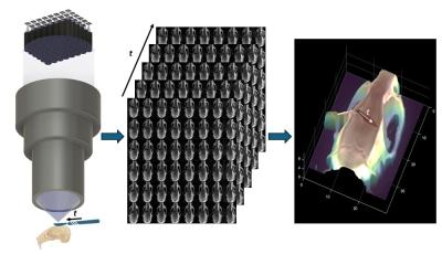
Light sheet microscopy is another technique that provides insight on the cellular level. Based on the so-called triangulation slice images of an object can be acquired and fused into a 3D image – or rather a 3D movie of live cells to be precise. These shorts show how the fusion of an egg cell and a sperm cell and the subsequent first cell division of the new embryo. Light sheet microscopy is used here primarily because these cells are highly light sensitive and thus would not survive conventional light microscopy.

“It can be really boring to sit at the microscope and look for something specific,” Tischer concedes. Therefore methods are being developed to automate the search. In automated feedback microscopy the software learns to detect and photograph interesting targets in a sample. This technique for example found the above-mentioned nuclei prior to mitosis. It is moreover an important tool in the Tara Oceans Project where over a period of three years the team aboard the French research schooner Tara collected water samples from all oceans on the globe. These were examined at EMBL for plankton – with sensational results: No less than 150,000 plankton species were discovered where previously the number of plankton species was estimated to be around 11,000. “For a human lab team this would have been a task for a lifetime,” Tischer laughs.
Profile:
Christian Tischer, PhD, studied physics in Heidelberg and received his doctorate from the European Molecular Biology Laboratory (EMBL), also in Heidelberg. Following post-doc work in biophysics in Amsterdam, he is currently at EMBL’s Advanced Light Microscopy Facility where he supports scientists with microscopy and image analysis.
29.05.2017