Image credit: Professor Hajime Monzen from Kindai University, Japan
News • Reusable and natural alternative
From sticky to solid: Ultrasound gel pad shows promise
Researchers assess the feasibility of a newly developed tamarind seed gum solid gel pad for clinical ultrasonography in human subjects
Ultrasonography is a non-invasive imaging technique used for real-time imaging. This versatile technique is used as a reliable diagnostic tool in various modalities. The conventional liquid gel used in ultrasonography is a critical component of this process that acts as an acoustic coupling medium, removing the air gap between the probe and skin surface. This allows improved image resolution and accurate diagnostic interpretations. However, the liquid gel has a quick drying time, which often compromises the image quality. Additionally, it may irritate patients and occasionally become contaminated. The clinician's performance is frequently impacted by the distinctive odor. To address these problems, researchers have been actively working on developing alternatives.
Against this backdrop, a team of researchers from Japan, led by Professor Hajime Monzen from the Department of Medical Physics at Kindai University, has developed a new solid gel pad made from tamarind seed gum with self-moisturizing property to solve these limitations and maintain the diagnostic precision. The research team included Dr. Takuya Uehara and Professor Yukinori Matsuo from Kindai University and MS. Megumi Ujifuku and Dr.Yutaka Watanabe from Kosei Clinic, Japan. The study was published in Scientific Reports.
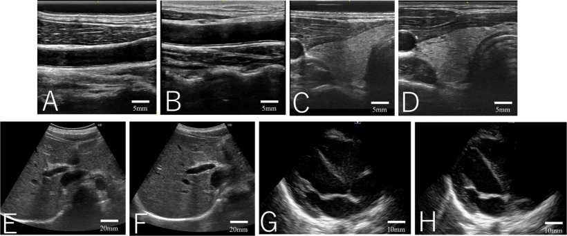
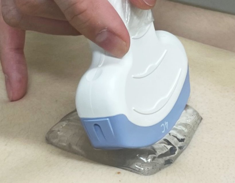
Image source: Uehara T, Monzen J, Ujifuki M, Matsuo Y, Watanabe Y, Scientific Reports 2026 (CC BY 4.0)
“Initially, I encountered a gel made only from water and tamarind. The developer explained that the continuous release of moisture was considered a drawback,” mentioned Prof. Monzen, while talking about the serendipitous origin of this innovative research. “However, I realized that this property could help prevent drying during ultrasound examinations, reduce the formation of air gaps between the probe and the skin, and thereby maintain stable image quality.”
The gel pad was developed using tamarind seed gum, a natural polysaccharide obtained by isolating and purifying tamarind seeds. Polyhydric alcohol and water were other two major components in the pad, along with preservatives being used to prevent microbial growth. Feasibility of this pad was tested on four healthy volunteers and multiple tissue types were targeted for imaging, for interpretability assessment.
In the future, this work may support ultrasound diagnostics that are easier to use, more patient-friendly, and more sustainable
Hajime Monzen
The thermal stability of the gel pad ensured its suitability for clinical applications. Its flexibility allowed it to conform to skin contours during ultrasound examinations while maintaining structural integrity. Continued syneresis of the gel allowed it to re-moisten, giving it a unique self-recovery capability. These properties, together, ensure optimal acoustic coupling and enhanced ultrasound image quality during ultrasonography.
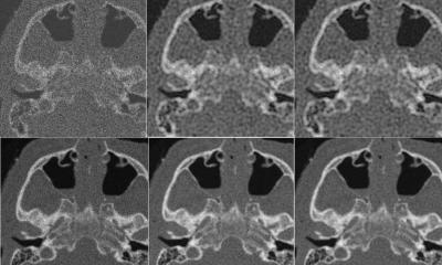
When compared directly with conventional liquid gel, the solid gel pad produced images of comparable quality across all examined sites. However, volunteer satisfaction was significantly higher when the solid gel was used. A major reason of patient discomfort is associated with the adherence of conventional liquid gel to chest hair and the difficulty of removal. The solid gel did not adhere to body hair and could be removed easily, resulting in reduced patient discomfort.
The use of this gel pad is expected to improve patient comfort and satisfaction during ultrasound examinations and ultrasound-guided procedures in regular clinical settings. This improvement may encourage patients to undergo examinations more readily, increasing chances of early diagnosis and timely treatment. It can also reduce operational costs related to gel heating and post-examination cleaning, making it accessible for patients from diverse economic backgrounds.
This gel pad can be stored at room temperature and is easy to handle, expanding its potential beyond routine clinical settings. It can be used for emergency medicine, disaster response, and medical practice in resource-limited settings.
Taken together, this solid gel pad is expected to generate meaningful social and economic benefits over the next five to ten years.
Prof. Monzen concludes by highlighting the long-term impact of this research, “From an academic perspective, this study helps clarify how the properties of tamarind seed gum relate to the way ultrasound waves travel. It presents a new approach to designing materials used in ultrasound examinations. In the future, this work may support ultrasound diagnostics that are easier to use, more patient-friendly, and more sustainable.”
Source: Kindai University
04.02.2026