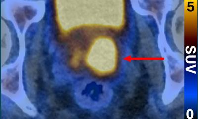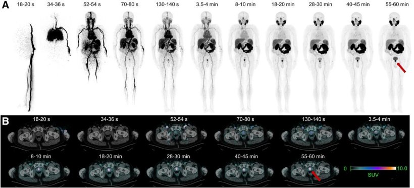
Dynamic total-body PSMA PET. 76-year-old patient with prostate cancer (PCa) (PI-RADS 2; initial PSA, 19.7; PSA density, 0.82). Representative reconstructed dynamic total-body PET maximum-intensity projection images (A) and transaxial fused PET/CT images (B). Red arrows indicate area of intense uptake in prostate (PRIMARY score 5; ISUP 4 after biopsy; SUVmax, 55.6).
Image source: Farolfi A, Sgro CM, Brusa I et al., Journal of Nuclear Medicine September 2026 (CC BY 4.0)
News • Combined approach
Prostate cancer: PSMA PET/CT-targeted biopsy improves detection
PSMA PET/CT-targeted biopsy detects more clinically significant prostate cancer than systematic biopsy alone in men whose MRI findings are negative or inconclusive yet have persistent clinical risk factors.
According to new research published in the Journal of Nuclear Medicine, combining the two biopsy approaches is even more effective, nearly doubling the detection rate. This strategy may help physicians identify clinically significant cancers that would otherwise be difficult to detect, while potentially avoiding unnecessary procedures in appropriately selected patients.
A negative or unclear MRI does not completely rule out clinically significant prostate cancer. As a result, some men may undergo repeated or extensive biopsies, while relevant tumors may still be missed
Andrea Farolfi
Prostate cancer diagnosis typically involves prostate biopsy using a systematic (sampling from a pre-defined area) or image-guided approach. Multiparametric MRI is considered the cornerstone for detecting and localizing clinically significant prostate cancer, enabling targeted biopsy and reducing overdiagnosis of slow-growing disease. However, up to 20%–30% of clinically significant prostate cancer cases may be missed in patients with negative or inconclusive MRI findings.
“A negative or unclear MRI does not completely rule out clinically significant prostate cancer. As a result, some men may undergo repeated or extensive biopsies, while relevant tumors may still be missed,” said Andrea Farolfi, MD, nuclear medicine physician at the Nuclear Medicine Unit, IRCCS Azienda Ospedaliero-Universitaria di Bologna, Bologna, Italy. “Determining whether PSMA PET can refine risk stratification and guide targeted sampling for these men could have a direct impact on clinical decision-making.”
In the prospective, single-center study, 63 men with negative or inconclusive MRI scans underwent 68Ga-PSMA-11 PET/CT followed by PET fusion-guided prostate biopsy. A subset of 11 patients underwent dynamic total-body PET. Researchers assessed the diagnostic performance of PSMA PET/CT–guided targeted biopsy versus systematic biopsy and explored the value of semiquantitative and dynamic total-body PET parameters in predicting clinically significant prostate cancer.
PSMA PET/CT was positive in 25 of the 26 men who were ultimately diagnosed with clinically significant prostate cancer. PSMA PET/CT-targeted biopsy detected clinically significant cancer in 37% of the study population, compared with 21% using systematic biopsy alone. Combining PSMA PET/CT-targeted biopsy with systematic biopsy increased the clinically significant prostate cancer detection rate to 41%. SUVmax was independently associated with clinically significant disease, and exploratory dynamic total-body PET demonstrated distinct kinetic patterns in aggressive lesions, particularly in unclear cases.
“Our findings suggest that PSMA PET/CT could provide an additional tool for identifying men who remain at risk despite non-diagnostic MRI findings and for directing the biopsy toward the most suspicious area,” said Farolfi. “If confirmed in larger studies, this approach could make the diagnostic pathway more accurate and personalized.”
Source: Society of Nuclear Medicine and Molecular Imaging
07.10.2026