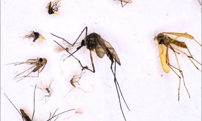
Source: Centers for Disease Control and Prevention (CDC)/Dr. Libero Ajello
Article • Travel medicine
Parasites & company – the radiologists' view
Sunburn and happy memories are not the only things we can bring home from a holiday. Sometimes parasites, fungi, viruses or bacteria from distant countries accompany our return, later to become noticeable in unpleasant ways, often to pose a real health threat. At the German Radiology Congress in Leipzig, Dr André Lollert and colleagues ventured into the world of tropical and travel medicine. The senior paediatric radiology consultant at the University Medical Centre of the Johannes Gutenberg University, in Mainz, provided an overview on which of these ‘stowaways’ can also enter German hospitals – and how radiology deals with them.
Report: Wolfgang Behrends

In his Leipzig presentation, Dr André Lollert spoke about worms (helminths), such as Echinococcus and Schistosoma; fungi of the genus Histoplasma, and about viral and bacterial infections. These pathogens mostly enter the body by ingestion (less frequently via skin injuries) and cause a wide range of diseases. ‘It’s mainly increasing tourism, but also migration that bring into our hospitals diseases whose origins lie in very different parts of the world. Therefore, the probability of being confronted with these exotic diseases in clinical routine is rising.’ Whilst native parasites such as fox or dog tapeworms are relatively common, the occurrence of melioidosis, a dangerous bacterial infection which is mainly found in Southeast Asia, is also on the increase.
If patients come to hospital after being abroad, these cases often can be quite harmless – the classic being travellers’ diarrhoea. ‘However, depending on the country the patient visited, a more exotic, differential diagnosis must be considered.’ For people whose immune defence is impaired by age or illness these pathogens can be life threatening. ‘Worms, in particular, move through the body once they have entered it, so different organs can be affected,’ Lollert explained.
An image alone is not enough
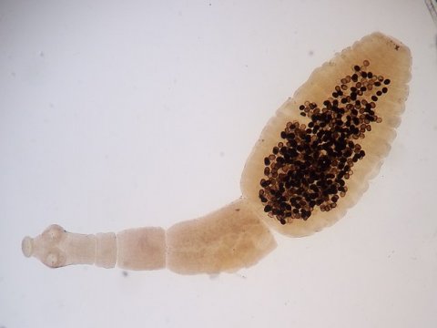
For the radiologist, the pathogens frequently remain invisible – only the traces they leave in the body can be seen. ‘Each parasite has a preferred part of the body where they settle. Whilst Echinococci often spread in the liver, histoplasmosis or melioidosis usually affect the lungs.’
Since the diseases may manifest in a very typical way, the interaction between radiology and patient anamnesis is vital, says Lollert: ‘Imaging alone does not deliver a reliable diagnosis. The region a patient visited provides important clues as to which pathogens could be the cause.’ If necessary, patho-logy or microbiology can provide confirmation via biopsy.

Source: Dr. André Lollert
Talking to the patient, as well as taking the patient’s symptoms into account, can also indicate which imaging procedures should be used. ‘If an Echinococcosis with neurological symptoms is suspected, the procedure of choice would be an MRI scan of the brain. In the abdominal region, the typical liver cysts that develop after a parasitic infestation can be visualised with ultrasound. The imaging modalities are as diverse as the possible pathogens.’
Radiological findings also affect treatment. Whilst some pathogens can be surgically removed, in other cases patients are treated with antibiotics or antihelminthics. Imaging procedures can also be used to monitor the effectiveness of this treatment.
Climate change drives vectors north
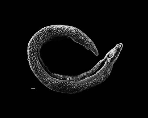
David Williams, Illinois State University, Schistosoma 20041-300, marked as public domain, details at Wikimedia Commons
Along with travel, other factors also spread exotic diseases, Lollert added. ‘Global climate change makes it possible for certain types of mosquito to move further north. These insects often transmit diseases that have, until recently, hardly been found in Europe.’ As yet, there have not been any significant clusters of these vector-borne diseases in Germany; but, he said, the transmission vectors could become more relevant in the future.
Profile:
Dr André Lollert is a senior consultant in paediatric radiology as well as radiation safety officer at the University Medical Centre of the Johannes Gutenberg University in Mainz. Whilst his research focus is on paediatric radiology he also specialises in the diagnosis of metabolic and infectious diseases. In 2017, Lollert was awarded the Publication Prize of the Society of Paediatric Radiology for his cancer research work in the field of quantitative imaging procedures.
08.07.2019


