Cardiac MRI
How does that work again?
You are curious to know what this cardiac MRI thing is all about? You want to brush up on your cardiac MRI knowledge? Then we are afraid you have to delve into the technical basics. Sounds boring? It sure isn’t, says Dr Harald Quick.

The Professor of High-Field and Hybrid MRI and Director of the Erwin L Hahn Institute for MRI at the University Duisburg- Essen in Germany is known for his entertaining introductions into the important technical foundations of MRI.
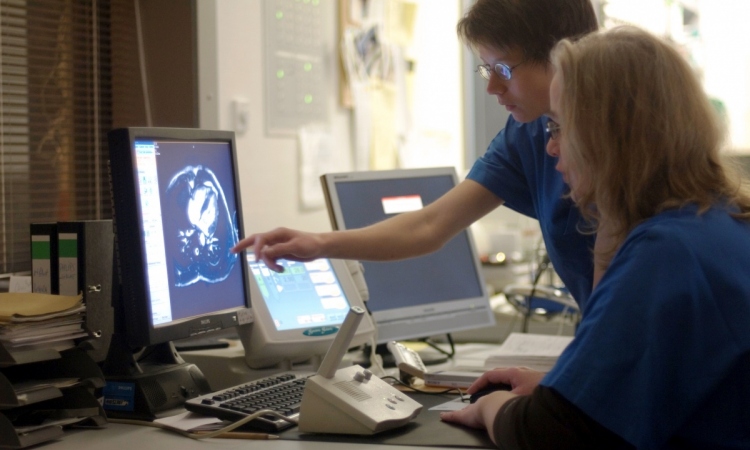
Dr Quick focuses on basic questions which are pertinent to every-day work with the MRI scanner such as the requirements high-frequency coils have to fulfil in order to be able to deliver excellent image quality or why cardiac MRI scans have to be ECG-triggered. “Radiologists are well advised to familiarize themselves with the basics of cardiac MRI and not to leave the settings to their technicians. After all, they are the one to decide which questions have to be answered and what needs to be visible on the images,” recommends Harald Quick.
ECG triggering, for example, controls the timing of the scanner’s image acquisition; it ensures that certain sections of the image are always acquired in a certain phase of the cardiac cycle. This enables extremely sharp images and allows the radiologist to evaluate the cardiac rhythm or to look at short film sequences that show the heart slice by slice. “This,” says Dr Quick, “Is the foundation of the evaluation of cardiac function. But you can also look at the blood volume in the left ventricle when the heart is fully dilated or when it has entirely ejected the blood. Thus you arrive at parameters such as ejection fraction and cardiac volume.”
The heart in slices
The major advantage of MRI is the fact that not only axial and transversal images can be acquired but that they can be produced from any angle and direction. “The ability to image in any plane however makes the selection of the appropriate settings more complex. While the heart is slightly tilted, imaging normally aims at standard orientations, thus the radiologist must ensure that the correct angle is selected,” MRI expert recommends.
An important standard view is the four-chamber view which displays all four chambers and two of the four cardiac valves in one plane which allows the assessment of valve function and tightness. Jets, for example, that is small signal losses, indicate turbulences caused by regurgitation,” explains Harald Quick.
“And last but not least parallel imaging techniques are routinely used in cardiac MRI today”. These techniques do not reduce the actual examination time but increase the speed of image acquisition and thus provides more and sharper images – which can significantly facilitate the diagnosis of this moving organ.
PROFILE:
In February 2014, Professor Dr Harald H. Quick, who is an engineer by training, was appointed Professor of High-Field and Hybrid MRI and Director of the Erwin L Hahn Institute for MRI at the University Duisburg-Essen in Germany. He gained professional experience inter alia as research associate at the Department of Radiology at the University Hospital Zurich, Switzerland, and at Johns Hopkins University in Baltimore, USA. From 2009 to 2014 Harald Quick was Professor of MRI and Deputy Director of the Institute of Medical Physics at the University of Erlangen, Germany.
29.11.2014