Handwriting revisited
Annick Chapoy reports
A specialised brain area involved in the production of written language was first empirically described by 19th century scientist S. Exner. At that time, the only way to investigate was by post mortem study of patients who had experienced writing problems during their lives. Now, in France, a team of researchers led by Jean François Démonet, has applied state-of-the art technology to study handwriting.

Language is subdivided in two modalities, oral and written. Handwriting, unlike spoken language, is a recent invention of humanity and does not come naturally. In fact it requires a long learning phase. Consequently, the brain areas sub-serving writing are not naturally intended to fulfil these functions but become so through specialisation.
The Inserm (Institut national de la santé et de la recherche médicale) researchers in Toulouse used two methods to precisely identify this area not exceeding a few square millimeters. This tiny area is the very place where a piece of information goes from “abstract” (the word understood), to “concrete” the word with a written trace…
Unlike Exner who was observing actual lesions, the first approach consisted in producing temporary virtual lesions, or at least temporary lesions. Taking advantage of a neurosurgical procedure to treat benign brain tumours, a small electrode may inactivate very small portions of cortex through electric stimulation.
Patient volunteers remained conscious during this phase of the procedure and were able to perform several language tasks, including a dictation. But, as soon as the electrode made contact with a certain area, patients were completely unable to write letters, whereas they could still read and move their hands.
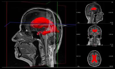
During the second approach, both right- and left-handed individuals were asked to perform the same tasks though this time during a functional MRI experiment . MRI or Magnetic Resonance Imaging makes it possible to visualise active areas in the brain by measuring changes in blood flow . Thanks to this technique, the activated areas in the brain are visualised in real time.
By comparing the areas activated during the different tasks it was possible to statistically define the existence of an area responsible for the production of written words and not, for example, naming aloud.
The importance of this progress really comes to the fore when the results obtained by these two methods are crossed. The team concordantly has managed to show that a small region in the upper part of the left frontal cortex is crucial for the written production of words. This area therefore allows the conversion of orthographical information (the letters that should be used) into graphic information (appropriate hand movements to write the letters in question). Indeed, words are abstract entities which must be “translated” in order to be made visible. A word may therefore be produced in different forms such as drawing, pronunciation or writing.
The team of Jean-François Démonet was able to pinpoint this “channel” in the brain that connects orthographic information to the movements of handwriting.
This study is a first step towards understanding other contemporary forms of writing such as the use of keyboards, but also writing disorders in diseases as varied as Parkinson's disease, vascular aphasia or developmental dyslexia .
25.01.2010