Sponsored • Advanced imaging solutions
Unlocking the Full Potential of Your Ultra High-Resolution and Photon Counting CT Systems
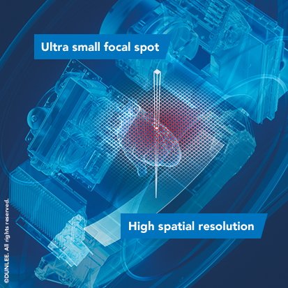
Today’s advanced CT applications require outstanding spatial resolution for both image quality and workflow. To make the most out of the current detector technology, attention has shifted to utilizing the X-ray source to increase spatial resolution, through extra small focal spots.
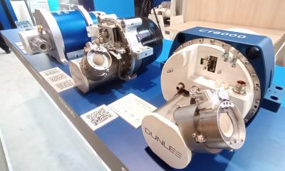
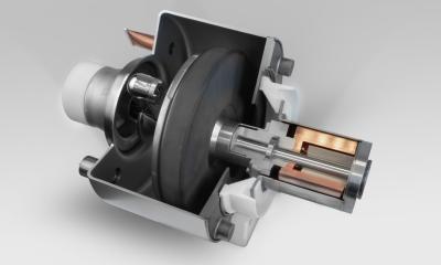
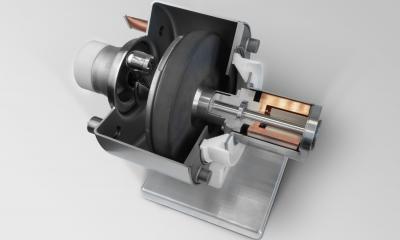
© Dunlee
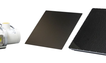
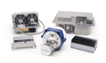
Dunlee’s extra small, stable focal spots are engineered to balance optimization of image quality, throughput, and heat management to help you get the most out of your ultra- high-resolution CT (UHR-CT) and photon counting CT (PCCT) systems.
The Rise of High-Resolution Imaging
CT imaging is expanding into applications that demand high spatial resolution to achieve images with sufficient detail for diagnostic confidence. Two leading approaches are shaping this trend: ultra- high-resolution CT (UHR-CT), which uses conventional energy integrating detectors (EIDs) with reduced pixel sizes for greater resolution, and photon-counting CT, which incorporates photon-counting detectors that offer higher resolution through smaller pixel sizes as well as spectral imaging capabilities.
These technological advances require equally advanced X-ray sources. Smaller detector pixels necessitate focal spots that are not only smaller, but also thermally and mechanically stable. In addition, high gantry rotation speeds for complex applications such as heart scans demand mechanically stable focal spots regardless of size. The extra small focal spots have been developed to address the stability needs of daily clinical operations.
Clinical Applications for High-Resolution CT Imaging
High-resolution CT enables precise visualization of fine structures. Applications include:
- Cardiology: For visualization of coronary arteries and soft plaques
- Oncology: For identification and characterization of small lesions and tumors
- Neurology: For detection of cerebral microbleeds and small infarcts and evaluation of fine neural structures
- Pulmonology: For detailed lung imaging and detecting interstitial changes
- Orthopedics: For visualization of spongy bone architecture, detection of microfractures, and assessment of implant placement.
Your Reliable Partner for UHR-CT and PCCT
Dunlee has over 100 years’ experience in developing, producing and integrating innovative components for imaging systems. It offers support during development and throughout the product lifecycle, contributing to its customers’ efficient production and go-to-market strategies. Visit www.dunlee.com to learn more.
To learn more about Dunlee’s approach to UHR-CT and PCCT, visit us at ECR 2026, Hall X2, Booth #204.
04.03.2026