Image source: Society of Nuclear Medicine and Molecular Imaging
News • After an initial negative scan
Treatment change for prostate cancer: study reveals benefit of second PSMA PET
Study findings further strengthen pivotal role of PSMA PET in the management of men with recurrence of prostate cancer after first-line therapy
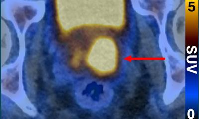
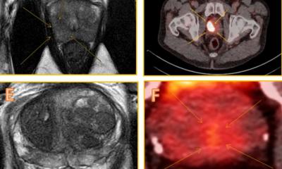
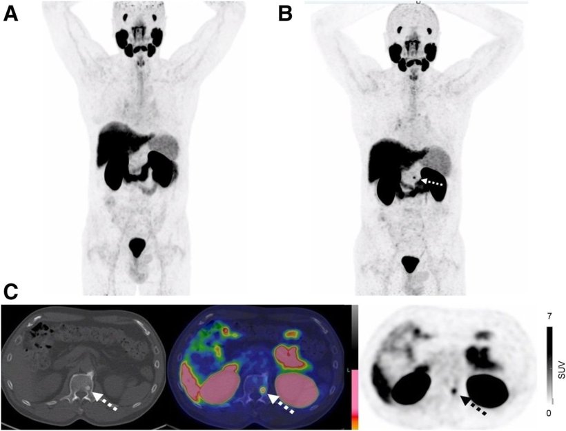
The findings were published in the Journal of Nuclear Medicine. Findings from the repeat PSMA scans, which included both local and distant disease, resulted in a change in management for nearly 50% of these patients.
Managing recurrent prostate cancer after first-line treatment, such as prostatectomy or radiation therapy, remains a clinical challenge. Although PSMA PET imaging has improved disease detection, 30% of patients still have no detectable disease on initial imaging, even as rising prostate-specific antigen (PSA) levels suggest recurrence. Few studies have examined whether repeating PSMA PET in this situation is worthwhile.
Understanding the extent of disease in patients who have initial negative PSMA PET scans provides valuable information for physicians as they create treatment plans
Ur Metser
“There is little information on the utility of repeating a PSMA PET after an initial negative scan,” said Ur Metser, BSc, MD, FRCPC, professor of radiology at the University of Toronto and head of the Division of Molecular Imaging at the Joint Department of Medical Imaging at Princess Margaret Cancer Centre in Toronto. “In our study, my colleagues and I sought to determine the benefit of a second PSMA PET scan, as well as to assess predictors for positive PSMA PET scans.”
The study included 210 patients from the Registry for Recurrent Prostate Cancer in Ontario who had more than one PSMA PET scan and whose initial scan was negative. The scan positivity rate, serum PSA, PSA doubling time, and management change after the second scan were compared with baseline data. Disease distribution was classified as local recurrence, locoregional, oligometastatic (fewer than five positive disease sites), or extensive metastatic (more than five positive disease sites).
A second PSMA PET scan revealed evidence of disease in 56% of recurrent prostate cancer patients who had an initial negative PSMA PET scan. A management change was necessary for nearly 50% of patients, in particular those with oligometastatic disease. Researchers found that repeat imaging was most likely to detect disease in patients with higher PSA levels and a PSA doubling time of less than twelve months.
“The findings in this study further strengthen the pivotal role of PSMA PET in the management of men with recurrence of prostate cancer after first-line therapy,” said Metser. “Understanding the extent of disease in patients who have initial negative PSMA PET scans provides valuable information for physicians as they create treatment plans.”
Source: Society of Nuclear Medicine and Molecular Imaging
13.07.2026