ESC Textbook of Cardiovascular Imaging now available
The idea of publishing a Textbook of Cardiovascular Imaging which accumulates the expertise of European cardiovascular imagers, is long awaited. The new textbook by the European Society of Cardiology is written with a patient rather than technology-based approach, helping clinicians to explain the condition to the patient, with the aid of imaging.

The book is divided into sections that treat specific themes involving theory and practice of cardiac imaging and its clinical use in all major cardiovascular diseases from coronary heart disease to cardiomyopathies.
" What is really different about the book is it is written by European specialists – 99% of contributors are from the European Society of Cardiology (ESC) and are key opinion leaders in their field. It is an ESC book so this implies quality. There are also videos and slides available so this makes learning easier," says Editor-in-Chief Professor J L Zamorano Gomez (president of the European Association of Echocardiography).
Imaging is an area that is rapidly developing in cardiology, explains Prof Zamorano, as it fulfils several important functions: “Imaging impacts in the diagnosis and the prognosis. Imaging helps in the monitoring of the treatment applied to the different patients. The key question is always how these modalities can help in our diagnosis. In the near future, CV Imaging will be multi-disciplinary."
The ESC became aware of the need for a book combining the accumulated expertise of European cardiovascular imagers. For those looking for a clear, easy-to-use textbook, the structure of this manual is very accessible, says Prof Zamorano:
“The textbook is split into two parts – the shorter gives the minimum knowledge needed to apply the related technique. The second part takes every single disease in cardiology and looks at how we approach these with the different modalities. The book is really disease orientated – we focus on the patient not the technology.
Imaging specialist Professor J Bax (Netherlands) is an editor of the textbook, who describes the expanding role of this technology, with greater use of imaging techniques in diagnostics and the consequent need for a textbook giving clear guidance:
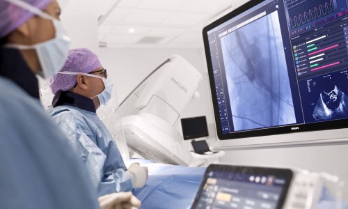
“Imaging in cardiology has increased dramatically and the number of procedures have increased, the number of techniques have increased, the requirement for training in imaging has increased, so imaging is very popular because people like to visualise things, as in daily life.
Most books are organised according to the four modalities: Echocardiography, Nuclear Imaging, Cardiac Computed Tomography (CT) and Cardiac Magnetic Resonance (MR) so I would call them technology driven. But at the end of the day most doctors have to take care of the patients so you want to know specific information about the disease and you can look in the introduction of this book to see which modality is appropriate.”
This book describes a new subspeciality that we will all need in the future: the imaging doctor, who can communicate with clinicians and help them resolve diagnostic dilemmas by choosing the correct technique and interpreting images using a patient-based approach.
Prof Bax explains: “Imaging is important because you want to preferably diagnose at an early stage and you do this easiest by visualising. If you can visualise, you can also determine the therapeutic approach. The basis is early diagnosis by visualisation guiding therapy.
The videos are extremely high quality using very modern technology and very good clinical case studies. They give a very clear illustration of what you see in a certain disease.”
The appropriate application of imaging procedures and accurate image interpretation are essential skills for the cardiologist. Each of the expertly written chapters in this textbook contains chosen images showing the full potential of modern cardiovascular imaging techniques.
Prof Bax adds: “If we take CT for example, this has developed from four into 20 row detector machines and that means it is now possible to capture the entire heart and arteries in one heartbeat. All modalities are developing very rapidly from two, to three to four dimensional.
On one hand, technology is developing rapidly and on the other there is a growing need to understand a patient’s condition and we do this by visualisation. When assessing the haemodynamic condition of a patient for example, seeing is understanding.”
The ESC CV Imaging textbook can be ordered here
08.01.2010


