Aired at EFORT: Guidelines for Charcot Foot
Meike Lerner reports
When Swiss orthopaedic surgeon Dr Marino Delmi, Past-President of the Swiss Foot & Ankle Society, Member of the Council of the European Foot & Ankle Society, and of the Scientific Foot & Ankle Council of the European Federation of National Associations of Orthopaedics and Traumatology (EFORT), met with other experts in the field, three critical diabetes topics were explored.

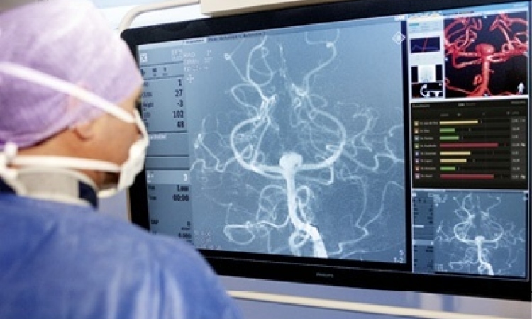
The first subject aired at the EFORT meeting was vascularisation and poor blood supply in diabetics -- and when to amputate, or not.
Vascularisation
‘We discussed what is necessary to maintain and spare the limb, especially the toe and so on, and if we have to amputate, the minimal amount we can carry out to maintain the most of the limb,’ said Dr Delmi. ‘With current techniques for limb sparing, surgeons amputate less and less. With the vascular status blood supply check we have an increasing number of good techniques and we also have some surgical techniques that can be used as vacuum therapy to help heal the scars.’
Diabetic Fractures
The second topic was diabetic fractures -- particularly of the ankle and foot. Problems include the vascular supply, neurological status, neurotrophic skin, nerves and bones, which are often osteoporotic. ‘It wasn’t clear that, after an ankle fracture in an ankle, the patient had a foot deformity, which is very tough to treat and could lead to amputation. It is very important to recognise the fact that the diabetic patient is not a normal fracture patient and must be treated with some specific techniques adapted to the patient, with some use of cement and specific screwing, and with more solid implants. We have to maintain the patient’s foot and keep him mobile for a longer time. All these could lead to a better evolution of the diabetic fracture, with less morbidity, wounding and infection, etc. If a surgeon is not aware of this, he can treat the diabetic patient with fractures in two ways, first as a normal patient with diabetic bones, or like a diabetic patient who is not normal and do no surgery, and so on. We should treat a diabetic patient with fractures as a normal patient, but with the postoperative care of the diabetic. Up to now this is not normal at all. So, surgeons first have to check what kind of patient they are treating.’
Their meeting and this discussion has lead to guidelines, he said: ‘The first is to recognise that a diabetic with a fracture is not the usual patient, so the bones and joints must be considered as for a normal patient but with specific, more solid implants, more solid devices, which produces better results.’
Charcot Foot
Charcot Foot is neuropathic but with a good vascularisation/blood supply, Dr Delmi pointed out. ‘We don’t know why; probably it’s a micro trauma with problems linked to neuropathy. We observe the full progress, the deformity or the foot with multiple bones fractures. Multiple dislocations of the joints can lead to deformity, which can be very important in the foot and ankle. It’s not a common problem but a very tough one. It can be seen in almost 10% of diabetic patients, from 1%-20% depending on the status. 10% is not a few, especially when you know that Charcot Foot can be very disabling if the foot cannot fit into a good shoe.
‘The surgeon or diabetic doctor, and especially the dermatologist, are must recognise this entity first -- I mean recognise fractures in a diabetic patient with a neuropathy and first treat the entity conservatively, but with special regard to this illness. If this doesn’t work you must take surgical measures and consider strong devices and strong implants. And, if you have a deformity you can stabilise or brace it.
The role of interventional radiology
‘MRI is important, particularly to check for the presence of an abscess of osteomalacia, and SPECT -- a mix of bone scanner and CT scan – enables us to see where the inflammation is active; this inflammation is linked with bone deformities. It is very important to be sure that the deformity is active, because you can have a deformity that is inactive, which means you don’t have to treat the deformity. However, if it is active, and the pain is coming from there, you’ll see that better with a SPECT than a CT scan.’
Interventional radiology includes a few methods to treat and revascularise diabetic foot. When it was suggested that this was only the role of the surgeon but has become more to do with radiologists, Dr Delmi pointed out that revascularising the diabetic foot is holistic – it involves the vascular surgeon, neurosurgeon, neurologist etc. ‘I’m sure that, for example within the amputation process, before amputation one of the ways to save or to spare the limb is for a vascular surgeon or radiologist -- using the new radiology techniques -- to increase blood supply to the foot.’
07.09.2010