News • Neonatology
Portable MRI in pediatric ECMO patients: promising premiere
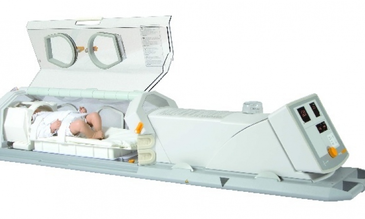
The neonatology team at the University Hospital Bonn (UKB) has conducted the world's first study of children receiving ECMO therapy using a mobile MRI scanner.
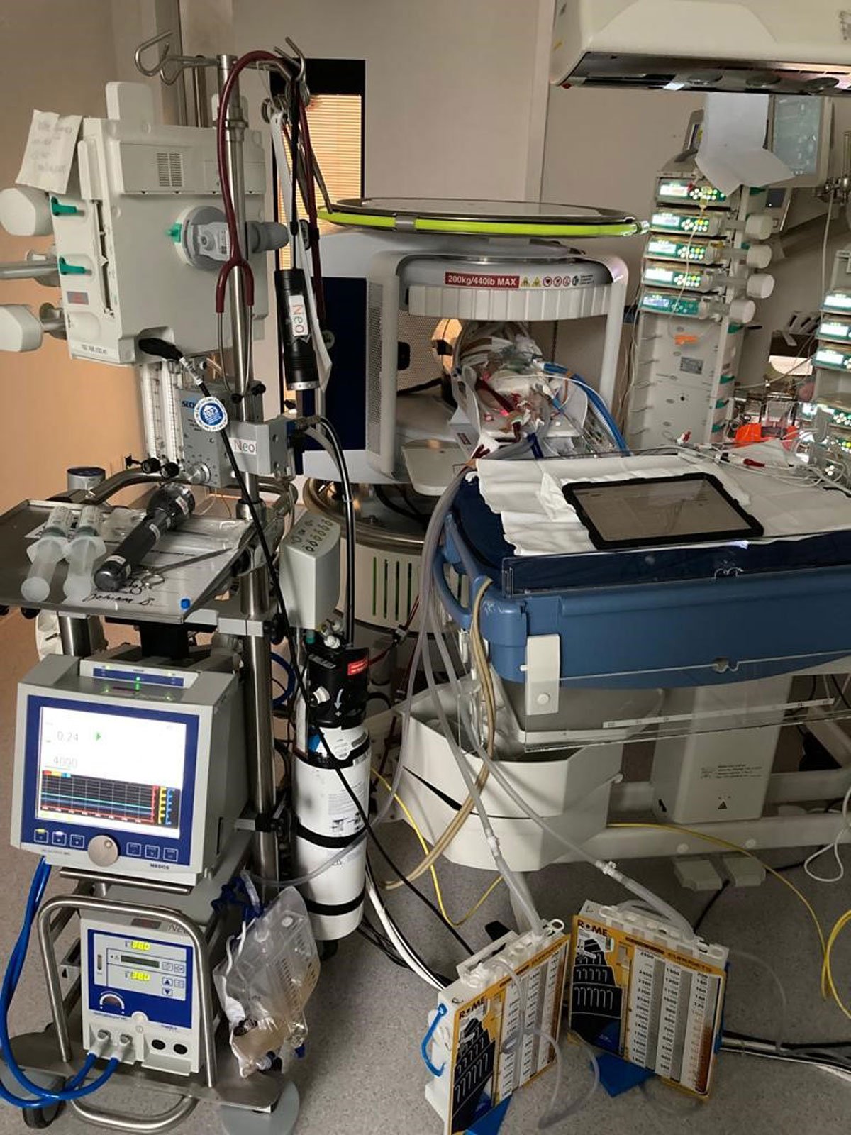
Image source: University Hospital Bonn/Prof. H. Sabir
The procedure, known as extracorporeal membrane oxygenation (ECMO), involves oxygenating the blood outside the body. The findings of the successful, innovative study of the first four pediatric ECMO patients using mobile MRI have now been published in the journal Critical Care.
Patients who require ECMO therapy beyond a conservative ventilator are critically ill. Reasons may include lung failure, heart failure or infection. Children who require this special procedure can only be treated at a special treatment center such as the Children's ECMO Center of the UKB, where they are closely monitored. Here, both newborns and older children are treated with ECMO therapy. "It is often necessary in this sensitive group of patients, even during ECMO therapy, to have an MRI of the brain to check the relevant structures in the brain. However, transport to a fixed device is unfortunately not possible," says Prof. Hemmen Sabir, who heads the department of experimental neonatology at UKB. Last August, a grant from the Bill Gates Foundation enabled him to purchase a mobile MRI, which is being used at the UKB for the first time in Germany to clinically test diagnostics on premature and newborn infants. So far, it has only been used for research purposes in London. The mobile MRI has already been in use at the UKB for more than half a year and represents a groundbreaking further optimization of diagnostics for neonatal patients.
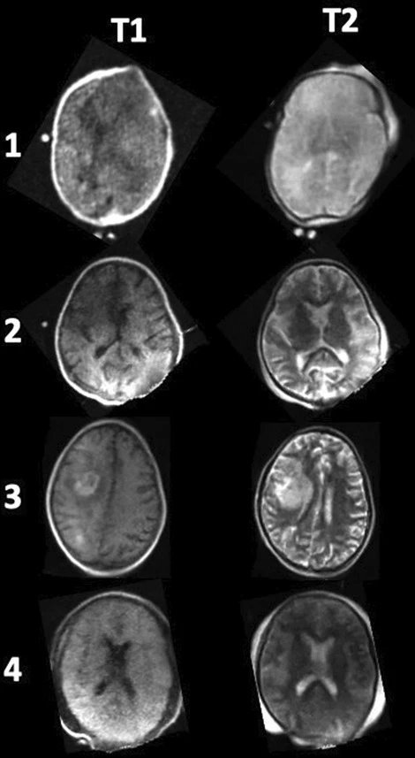
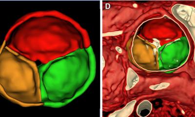
Image source: Sabir et al., Critical Care 2023 (CC BY 4.0)
25 children have since been scanned in the mobile MRI at the UKB - the youngest weighed only 450 grams, the oldest was already 10 years old. The mobile MRI was used for routine examinations and for further diagnosis of abnormalities, e.g. after asphyxia (oxygen deficiency at birth). To evaluate the image quality of the mobile low-field MRI, a comparison image was taken in the permanently installed normal-field MRI at the UKB for each of the children examined. "We were more than satisfied with the results. Although the image quality of the mobile MRI is not as high-resolution as that of a fixed device, the image data are ideal for emergency diagnostics and, above all, can be retrieved immediately. Among other things, we were able to detect brain hemorrhages, strokes or acute changes, such as the accumulation of cerebrospinal fluid, in the children examined so far and initiate the appropriate therapies immediately," says Prof. Sabir.
ECMO patients, however, pose a special challenge to the treatment team in connection with MRI diagnostics. "While adults have a tube inserted in the groin area to transport blood during ECMO therapy, children often have access at the neck. The patients have to be moved very carefully and the tube at the neck may only be moved minimally," explains Prof. Sabir. However, reliable diagnostics, for example of brain hemorrhages, is only possible with MRI imaging. The neonatology and pediatric intensive care team was therefore able to demonstrate in four pediatric ECMO patients that imaging using mobile MRI can be performed without any problems.
We obtained meaningful MRI images of the brain without changing the position of the neck cannula and without compromising the children's safety status
Andreas Müller
The patients studied were a newborn, a two-year-old, a nine-year-old and a ten-year-old child. One of the children was diagnosed with a major brain hemorrhage using the mobile MRI and was treated immediately. "The new findings prove that the scan can be performed safely. We obtained meaningful MRI images of the brain without changing the position of the neck cannula and without compromising the children's safety status. This represents an immense success for future MRI examinations of newborns and larger children who can only survive through the use of ECMO therapy," said Prof. Andreas Müller, Director of the Department of Neonatology at UKB.
Source: University Hospital Bonn
09.04.2023