GE launches a new monitoring technique
Working in tandem, two new technologies — SpiroDynamics and FRC INview — measure pressure and volume directly from a patient's lung.
During this system’s launch at this year’s Annual Congress of the European Society of Intensive Care Medicine in Barcelona, we asked Dr Ola Stenqvist, creator of SpiroDynamics and FRC INview, and Consultant Anaesthesiologist at the Department of Anaesthesia at Sahlgrenska University Hospital, Gothenberg, Sweden, what makes the system unique.
OS: SpiroDynamics enables clinicians to measure respiratory pressure directly from the patient instead of measuring it from the ventilator. Measurements are taken continuously in the patient’s airways, from where pressure down in the lung (where gas exchange takes place) is calculated. It’s like having a telescope in the air pipe to find out what’s happening in the lung. This tracheal pressure measurement has several clinical benefits: it is performed regardless of ventilator settings; it creates a dynostatic curve providing estimated alveolar pressure, and it offers enhanced detection of intrinsic PEEP due to ventilator settings or secretions.
FRC stands for Functional Residual Capacity and indicates the volume that is left in the lungs at the end of respiration. Even after respiration, normally there is a lung volume of two or three litres, which functions as a buffer. In acute lung injury (ALI) patients, this volume is decreasing significantly. Up to now, there have only been complicated systems to measure that volume – moreover, those systems were available for research purposes only, but not for every day clinical life. With FRC INview we can offer clinicians a tool to measure this volume at the bedside, with a 90% accuracy.
Since SpiroDynamics and FRC INview work parallel and continuously, and provide results directly at the bedside, we are talking not only about measurements but also about monitoring. This means clinicians receive all these data automatically – without interrupting the ventilation process.
What effect will this have on the treatment of ALI patients?
OS: On the one hand we can permanently examine the respiratory status – which of course means we can react much faster – and, on the other, we can look at changes in ventilator settings in real-time. For example, if a clinician tries to improve the lung volume by increasing pressure, he can see the results of his efforts immediately, without interrupting the ventilation to perform CT examinations.
By generating electronic snapshots in the ventilator, waves can be saved and compared with previous ones. Therefore, the course of the disease and changes caused by intervention can be monitored and analysed.
What have clinicians’ reactions been to these techniques?
OS: Interest among clinicians is huge because, although there has been a lot of research in respiratory mechanics, the measurement technology has been lacking. I’d say we rekindled the interest of physicians in this topic.
A completely new approach to mechanical ventilation
Since mechanical ventilation arrived over 40 years ago, ventilation therapy has involved adjusting airway pressure, flow and volume. However, when researchers in Toronto, Canada, developed the physiological concept of Neurally Adjusted Ventilatory Assist (NAVA), in which the patient’s own respiratory centre in the brain adjusts the ventilation pressure, the way in which patients suffering respiratory diseases can be treated radically changed.
Whereas conventional mechanical ventilators sense a patient’s effort to breathe, either by a drop in airway pressure or a reversal in flow – the last and slowest reacting step in the chain of respiratory events - NAVA senses electrical activity in the patient’s diaphragm – the earliest respiratory signal that can be detected.
Signals from respiratory control centre in the brain are transmitted through the phrenic nerve to the diaphragm, where a catheter captures the electrical activity of the diaphragm (Edi) and feeds it to the ventilator. NAVA responds by providing the requested level of ventilatory support to the patient. As the ventilator and diaphragm work with the same signal, the coupling between the two is virtually instantaneous.
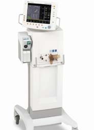
MAQUET Critical Care has added NAVA (Neurally Adjusted Ventilatory Assist) to its SERVO-i ventilator – a combination that presents a completely new approach for mechanical ventilation.
Potential benefits
• Improved patient/ventilator synchrony
• Lung protection by avoiding over or under assistance of a patient
• Enhanced patient comfort: improved synchrony helps minimise patient discomfort and agitation while it promotes spontaneous breathing
• The Edi signal can be used as decision support for medical staff regarding unloading or extubation
• The Edi signal can be used as a monitoring tool, providing data on
respiratory drive, volume requirements, effect of ventilatory settings and to gain indication for sedation and weaning.
The company points out that current SERVO-i users can upgrade their equipment with the NAVA function. For newly purchased SERVO-i, the only extra equipment needed is NAVA software, an Edi Module and an Edi catheter.
16.11.2006