Advancements in Telepathology
Professor Werner Schlake, President of the German professional association of pathologists, described teleconsultation as the most significant application of telepathology.
‘Teleconsultation means the professional exchange between pathologists,’ explained Professor Schlake. ‘Pathology is a very broad medical discipline, and therefore we have neither the financial nor the personnel resources to ensure that each pathologist is an expert in each aspect of the field. However, hospitals and physicians need the services of local, or at least regional, ‘general’ pathologists. In addition, we need access to specialist knowledge - for example, in lymphoma or liver diagnostics. If telepathology allows a regional pathologist to co-operate online with a reference or consultation centre for lymphomas, the entire range of pathology expert knowledge and services would become available even in remote areas. Therefore, it is crucial to establish a network of pathology expert knowledge. One of the most fascinating tasks of the German Professional Association of Pathologists is to promote and foster such a network.
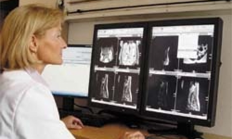
This is an ongoing process, because we have to adjust permanently to rapid technological developments. Until recently, telepathology was a rather limited application - we simply did not have the required computer storage capacities. The data volumes generated and processed during the digitisation of a single microscopic slide are many times larger than those of an X-ray image, which is basically due to colour and detail. In a pilot study in our institute we found that the average data volume of one microscopic slide is 800Mb - one microscopic slide! For certain diagnostic procedures such as breast cancer we need ten or twelve microscopic slides. However, today storage capacities have grown manifold, and at the same time prices have dropped significantly. Consequently, storage capacity is no longer the limiting factor it was a couple of years ago. The second important aspect is the scanner. The microscopic slide has to be scanned as a whole, or we have to take individual photographs at the microscope.
To help our members to begin telepathology the association installed a server, which, for personnel and financial reasons, is located at the Charité in Berlin. Currently, it is used primarily for the pilot project on mammography screening (see box). Every member of the association has access to this server, to ask for or render a second opinion. That means, at his microscope the pathologist creates a digital image of the specimen in question in different magnifications, and stores them on his hard drive. From there he downloads the images onto the server. Within a predefined time span, a competent colleague will render a second opinion. For us, this is the only way to explore the possibilities as well as the possible limitations of telediagnostics. How far can we go? Is the diagnosis based purely on digital material as good as the immediate microscope diagnosis? We don’t know for sure yet, but I assume it will be very close. Current technological development has shown that digitisation is the way of the future.
How is it guaranteed that, when downloaded to the server, the image quality is acceptable and comparable?
We provide guidelines concerning the technical minimum requirements, so the images will all be of decent and comparable quality.
Can all pathologists afford those technical requirements?
Definitely. There is just one issue that might pose a bit of a problem: the telephone lines. Universities usually have 100 megabit lines, but that is the exception. We try to achieve adequate results with 10 megabit lines. Even 2,5 megabit and DSL works - but then the data transfer does take a while.
Will the virtual microscope, a new development, play a significant role in the future?
Indeed, this is a fascinating development, which is still in its pilot stage. I worked with such a virtual microscope for three months. The exciting innovation is the scanning technology. This was developed by a working group in Budapest, and then Zeiss came on board and launched it under the name Miraxscan (see box). The microscopic slide that we usually see under the microscope is scanned. The brilliant innovation is the fact that one does not have to generate, store and transfer images of several microscopic slides but only one. And then I can enlarge this image on my computer, in the same way that I usually do under the microscope. That works wonderfully. This solves a problem that, until now, was considered insoluble. Moreover, this scanning technology works at an acceptable speed. Previously we needed about an hour to scan one microscopic slide. Now we need one to three minutes. You can prepare a whole set; it will be documented and marked with all the necessary data prior to the actual scan. In short: this will enable us to work on screen just as we do now under the microscope.
What is needed to be able to analyse digitised images with the virtual microscope?
Just like with any other teleconsultation project there are certain prerequisites needed: separate from the regular IT equipment a consultation server is required to up and download images. To download images you need a so-called image streaming software to enlarge the images. It works amazingly well. We tested it between the Charité and our institute in Gelsenkirchen. This scanning equipment is presently expensive, carrying a price tag of about 150,000 euros plus the storage devices.
One of the other great advantages of the virtual microscope is archiving. We are asked to archive every single microscopic slide for 10 years. These are glass objects, so we have to store tons of material. And we have to be able to retrieve every single slide quickly. Digital archives would make life much easier.
Could a digital archive really replace a physical one?
We are not really sure about that yet, but I have a hunch that, in the end, the digital archive will replace the physical one. Consider this: in an intricate procedure water is extracted from the specimen and replaced by paraffin. In the next step this solidified specimen is embedded in a larger paraffin block. Then very thin sections are cut off that block and stained. These slices are used for diagnosis. We are increasingly asked to also archive the ‘leftovers’ of the paraffin blocks from which we cut the sections. With a digital archive and a physical archive all possible questions that might turn up later can be answered. With a digitised archive alone that might not be possible. For example, many large institutes archive the paraffin block when cancer has been diagnosed. Recently, a new treatment for a certain kind of breast cancer was developed: if the tumour cells carry a certain amount of HER2/neu-proteins, an antibody therapy can be initiated. For the women concerned this is an interesting alternative, since with this antibody therapy a surgical intervention is no longer required. If a woman who has been diagnosed with breast cancer a few years back considers such a treatment, the physician first has to analyse the cells. To do so, he can examine the original paraffin block - if he archived it. If not, the woman has to undergo another biopsy. Therefore, we are asked to archive all blocks for 15 years. Digital archives - that would be the ideal solution.
Does digital archiving also open up new perspectives on quantitative evaluation of specimens?
Yes, with virtual microscopy we can, for example, quickly determine percentage distributions. A software-based system can tell me that, for example, 20% of the tumour cells are oestrogen positive, and 80% are negative. Today, we use more or less vague approaches for quantification. We can also perform analyses using a proliferation marker, which tells us what percentage of cells are in growth stage. We can find out whether a tumour grows fast or slow. Here, digitisation offers many possibilities.
So, is this the end of the traditional microscope as we know it?
I think that the digital procedure will replace the microscope for certain routine diagnostics. When we did the pilot study in our institute we found that digitisation offers a number of possibilities that the traditional microscope can’t provide. For example, I can display on screen an entire overview of several specimens on one screen - extremely important in some cases. Or if one has differently stained tumour sections, you can view and compare all of them on one computer screen. I can’t do that with the microscope. Therefore I believe that digital microscopy will replace certain core tasks of the microscope. But first we must prove that the digital procedure provides the same level of quality as the manual one.
Is frozen section an issue in telepathology?
Yes, this has a number of basic problems: first, it is performed by non-pathologists. Tactile sensitivity is very important in a pathologist’s work, so it’s an integral part of his training. The eye must be trained as well. Consequently, the tactile sensitivity and eye of a pathologist are quite different from that of a surgeon. Second: the staining procedure of a frozen section is limited. The frozen section does not allow the sophisticated technical procedures a paraffin block can undergo. Third: the number of intra-operative frozen sections, e.g. in mammography, is declining rapidly because breast cancer prevention and diagnostics start at a much earlier age. Consequently, malignant tissue changes are also detected earlier. If a physician suspects a tumour that he can neither see nor feel, he can perform a frozen section - but only of one or two tiny areas. The selection of these areas is random. Maybe these frozen sections do not contain any important findings, but when later the tissue is examined more closely, the pathologist does find suspicious foci, then you have to tell the patient, sorry, we made a mistake in our initial diagnosis. That is very frustrating. Therefore, frozen sections are today no longer an option with tiny lesions. Consequently, frozen section is currently no issue for telepathology. We’d rather focus on the really innovative potential of telepathology: teleconsultation. It is cheaper than the traditional method of sending out a specimen by mail or courier, and therefore an attractive option in terms of costs. Not to mention the fact that it is much faster and easier to handle. However, most importantly, teleconsultation offers the pathologist - wherever he is - access to specialist knowledge, and this means that the patient, physician and local pathologist can benefit from state-of-the-art diagnostics.
08.08.2006