News • ESR
Foundation of new subspecialty society for hybrid imaging
The European Society of Radiology (ESR) is proud to announce the foundation of a new subspecialty society, the European Society for Hybrid Medical Imaging (ESHI).


The European Society of Radiology (ESR) is proud to announce the foundation of a new subspecialty society, the European Society for Hybrid Medical Imaging (ESHI).
PET/MR has long been studied for oncology but the technique also holds promise in cardiovascular applications, according to a panel of experts at the recent International Conference on Nuclear Cardiology and Cardiac CT (ICNCT).

The vast amounts of data accumulating in breast diagnostics require new methods to extract clinical information in a practical way. When dealing with large amounts of data that is too big or too complex to be analysed with traditional data processing applications, the talk today is of ‘Big Data’. The data volume accumulating in breast diagnostics has become increasingly complex over recent…

The view across the Atlantic – it fills Professor Fabian Kiessling, Chair of Experimental Molecular Imaging at the RWTH Aachen (Rhine-Westphalia Institute of Technology Aachen), with optimism. The USA offers more opportunities for molecular imaging. Only recently, new tracers for Alzheimer’s were accepted as reimbursable in some centres, whilst the development of new diagnostics in Europe…
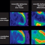
Dr Gerald Antoch, professor of radiology and chairman of the department of diagnostic and interventional radiology at Düsseldorf University Hospital and active member of several scientific societies, delivered the prestigious Wilhelm Conrad Röntgen Honorary Lecture at ECR 2015 on ‘Hybrid imaging: Let the two worlds of radiology and nuclear medicine come together’. Report: Marcel Rasch
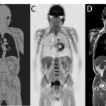
PET/MRI scanners have great potential because they combine the strengths of two different systems. Previous problems resulting from respective, mutually exclusive physical effects of both procedures have been resolved. Now these scanners are being introduced to the hospital and assist in the detection of the position and spread of tumours as well as their metabolic activity, says Dr Harald H…

The ‘MRI of the Adnexa, Essentials and Beyond’ session from at the MR 2015 symposium held in Garmisch, Austria, presented tips and tricks in clinical routine before moving to research that combines imaging and genomic data to better evaluate ovarian cancer. Report: John Brosky

Hybrid imaging is of little clinical value. PET-MRI, according to many experts, is the best clinical procedure to confirm coronary heart disease (CHD). Report: Axel Viola
Someone once described the fusion of positron emission tomography (PET) and magnetic resonance imaging (MRI) as a great technology looking for an application. Report: John Brosky
Approach could improve treatment of drug-resistant infections. Combining a PET scanner with a new chemical tracer that selectively tags specific types of bacteria, Johns Hopkins researchers - working with mice report - have devised a way to detect and monitor in real time infections with a class of dangerous Gram-negative bacteria.
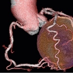
The significant benefits of cardiac catherisation remain undisputed. However, cross-sectional imaging modalities are serious competitors when it comes to arriving at the right diagnosis.
European Hospital met up with Professor Harald H. Quick, PhD, who was appointed Director of the Erwin L. Hahn Institute (ELH) for Magnetic Resonance (MR) Imaging this February.
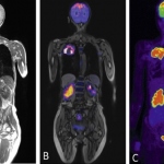
The German Cancer Research Center (Deutsches Krebsforschungszentrum, DKFZ) is sending a promising duo into the race against cancer: A new PET/MR system that can combine high-resolution images with functional information to improve cancer diagnosis.

Modern imaging techniques greatly enhance the treatment selection

Hedvig Hricak, Chair of the Radiology Department at the Memorial Sloan-Kettering Cancer Center, New York, USA, describes emerging applications and potential trends in gynaecological cancer treatment described at the 15th International Symposium Crossing Barriers

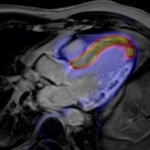
MRI has become the gold standard for many indications in cardiac imaging, apart from imaging the coronary arteries. For function and morphology assessment, MRI is the leading technology. A further advance into as yet unknown territory is myocardial imaging aided by one of the first integrated 3-Tesla PET/MR systems currently used at the Institute of Radiology, Essen University Hospital,…
Experts across Europe believe the combination is beginning to demonstrate its broad potential as a hybrid imaging tool
UK researchers are working on a new MRI technique called hyperpolarised MRI – or Dynamic Nuclear Polarisation (DNP) – that can utilise more of the available nuclei than traditional MRI, helping to overcome some of its limitations by increasing sensitivity 10,000-fold or more. DNP is part of a longer-term aim to improve cancer mortality with the help of novel cancer imaging tools.

When asked about his vision of imaging in the year 2020, Professor Bernd Hamm MD, director of the three radiology clinics at the Charité University Hospital in Berlin, qualified his focus: ‘Technology is always only a vehicle. When we talk about road traffic, we don’t talk about the design of cars but about structural issues’

Virtual FDG-PET/CT bronchoscopy has been found to be a technically feasible tool for the detection of lymph node metastases in non-small cell lung cancer patients with good diagnostic accuracy, according to researchers at the Department of Diagnostic and Interventional Radiology, University Hospital Dusseldorf and Essen.

Quick, simple and comfortable – the RADbook 2012 on your iPad. Download the RADbook app free of charge today and have all state-of-the-art diagnostic imaging systems at your fingertips.

A series of papers presented at the European Congress of Radiology on Friday have highlighted how hybrid imaging is helping radiologists achieve better results in the diagnosis of patients’ conditions. In a session focussing on molecular imaging and entitled “Hybrid imaging: PET-CT and MR-PET”, findings from ten different research papers were detailed by radiologists from Italy,…
Radiology constantly evolves. There are technical advances in terms of the capabilities of various modalities, greater clarity from contrast agents that are also safer for patients, and innovation in techniques that gains even greater performance from existing equipment, or enables further development.
Royal Philips Electronics is announcing 510(k) clearance from the Food and Drug Administration (FDA) for the company’s first commercially available whole body positron emission tomography/magnetic resonance (PET/MR) imaging system, the Ingenuity TF PET/MR.
Although like a conventional MR scanner the unassuming exterior is misleading. The casing houses a powerful interior. This is the new Siemens Biograph mMR, a hybrid that contains a specially developed PET component fully protected against magnetic field interference.