


News • Study confirms association
CT scans in young people increase cancer risk
A multinational study confirms a strong and clear association between exposure to radiation from CT scans in young people and an increased risk of blood cancers.


Since its introduction in the 1970s, computed tomography has been a mainstay of radiology. Its overlay-free representation of body structures and the rapid availability of images make CT indispensable in the diagnostic assessment of numerous diseases, especially in emergency medicine. Modern CT systems not only offer innovative procedures for better image quality, but also reduce radiation exposure.
A multinational study confirms a strong and clear association between exposure to radiation from CT scans in young people and an increased risk of blood cancers.
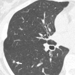
Diagnosing early-stage lung cancer with low-dose CT screening dramatically improves the long-term survival rate of cancer patients, a large-scale, 20-year international study shows.
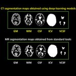
A new AI method for CT brain imaging may bring the modality to the level of detail usually reserved for MRI scans. This could enhance diagnostic support for conditions such as Alzheimer's disease.

Radiologists called for action to reduce the release of contrast media in the hospital’s wastewater after contrast-enhanced examinations in a dedicated session at ECR 2023.
Identifying victims of major disasters remains a significant challenge for investigators. Often, identification can take weeks or longer but new approaches are paving the way for greater accuracy and quicker identification whilst preserving the body without unnecessary invasive investigation. An expert session at ECR heard about how new imaging technology can help with disaster victim…

While screening programs for several of the commonest cancers are now well established, lung cancer screening has yet to reach anywhere near the same proportion of at-risk patients.
AI can use data from low-dose CT scans of the lungs to improve risk prediction for death from lung cancer, cardiovascular disease and other causes, a new study finds.
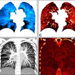
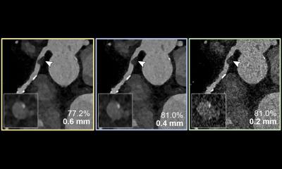
Photon-counting CT allows for a comprehensive, simultaneous evaluation of lung structure and function, something not possible with standard CT, according to a new study.

Should radiation dose from previous medical imaging be considered when a new scan is made? UK experts issued a joint position statement that favors clinical need over prior exposures.

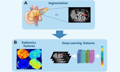
New research shows how AI can be used to fuse images from clinical X-ray CT and MRI scans to allow a clearer and more clinically useful interpretation of the images.
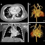
Photon-counting CT enables accurate diagnosis of coronary artery disease in high-risk patients, a potentially significant benefit for people previously ineligible for noninvasive screening.

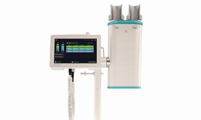
Iodinated contrast media (ICM) enhance CT imaging, but its single-dose packaging is increasingly proving at odds with modern, more sustainable imaging practices. New award-winning research by a radiology resident and faculty members at Vanderbilt University Medical Center in Nashville, Tennessee, proposes a promising alternative: A switch from using single-dose injectable contrast media kits to…

Transient ischemic attack (TIA) emergency department (ED) encounters with incomplete neurovascular imaging were associated with higher odds of subsequent stroke within 90 days, a new study finds.
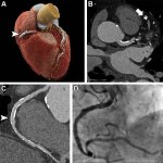
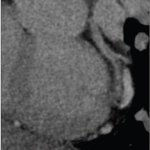
A new advanced form of CT imaging offers better cardiovascular imaging quality compared to dual-source CT (DSCT) in infants with suspected cardiac heart defects, according to a new study.
When is a CT scan justified, i.e. when do the benefits of a CT scan for the patient outweigh possible risks associated with radiation? Justification has been a major issue among radiologists ever since CT has become widely available and widely used. With regard to dose the answer is the well-known ALARA principle: “As low as reasonable achievable“. Now, the European coordinated action on…

Dunlee unveiled its new oncology bundles onsite at ECR. The bundles combine components that have been tested and verified to work together so clinicians can offer state-of-the-art onboard Cone Beam CT (CBCT) in facilities.

It’s clear that radiology is lacking in the “green” department: healthcare still causes a large share of global greenhouse gas (GHG) emissions, not least due to diagnostic imaging. Dr Sarah Sheard from Imperial College Healthcare, UK, invited her ECR audience to take a closer look at radiology’s climate footprint – and revealed ways to make the field more sustainable.

Radiology practitioners have highlighted the benefits of cone beam CT in delivering high resolution at a low dose. Delegates at ECR 2023 in Vienna heard how cone beam CT (CBCT) could replace multidetector CT (MDCT) in some areas and is already showing cost-effectiveness benefits.
According to an accepted manuscript published in the American Journal of Roentgenology (AJR), using a dual-source CT (DSCT) scanner for coronary CTA can facilitate clinical processes by eliminating the need to administer beta-blockers for heart rate control while decreasing nondiagnostic examinations.