© CEA
News • High-resolution brain imaging
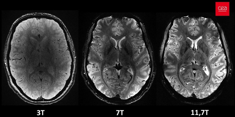
11.7 Tesla: First images from the world's most powerful MRI scanner
The French Alternative Energies and Atomic Energy Commission (CEA) is revealing a series of in vivo human brain images acquired with the Iseult MRI machine and its unmatched 11.7 teslas magnetic field strength.
This success is the fruit of more than 20 years of R&D as part of the Iseult project, with one pillar goal being to design and build the world’s most powerful MRI machine. Its ambition is to study healthy and diseased human brains with an unprecedented resolution, allowing the discovery of new details relating to the brain’s anatomy, connections, and activity.
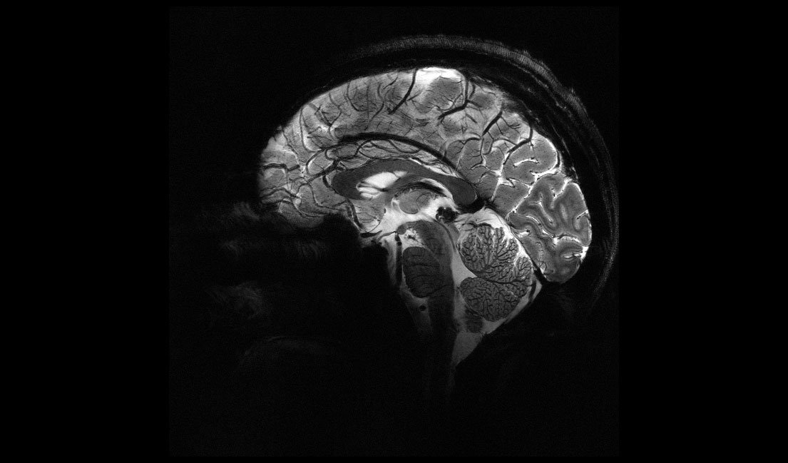
Just about four minutes. That’s all it took to acquire some of the most remarkable anatomical images of the brain from participants in the first study involving the Iseult MRI machine. The scanner, which uses magnetic resonance imaging technology, has a magnetic field intensity of 11.7 teslas, making it the most powerful in the world. The images have an impressive resolution for such a short acquisition time – 0.2 mm in-plane resolution and 1 mm slice thickness, which represents a volume equivalent to a few thousand neurons. For comparison, the same image quality would require hours with MRI scanners currently available in hospitals (1.5 or 3 teslas). This is not realistic in practice as patients would not be comfortable and any movement would “blur” the image.
© CEA
Achieving such detailed resolutions will allow researchers to obtain previously unattainable information about brain mechanisms, understand how our brain encodes our mental representations and find out what neuronal signatures are associated with the state of consciousness.
The level of detail achieved with the Iseult MRI machine will have an impact on medical research. Firstly, the ultra-detailed anatomical information will support diagnostic and health care for neurodegenerative diseases such as Alzheimer’s and Parkinson’s. Secondly, the Iseult MRI machine will facilitate the detection of some chemical species with weak signals that are hard to capture at lower magnetic fields, such as:
- lithium, a drug used to treat bipolar disorders; making it possible to precisely assess its distribution in the brain and better understand its efficacy;
- molecules actively involved in brain metabolism, such as glucose and glutamate; such information will directly contribute to the characterisation of many brain diseases (gliomas, neurodegeneration, etc.)
Neuroscientists, physicists, mathematicians and physicians thus worked together to develop the tools and models that will help better understand how healthy and diseased brains work, expanding the horizons of explorations on the human brain
Anne-Isabelle Etienvre
"With the Iseult project, a whole new world is opening up before our eyes, and we are excited to explore it. We still need several years of research to develop and improve our acquisition methods and ensure that the data has the highest quality possible. Our goal is to investigate neurodegenerative diseases by 2026-2030, as well as other diseases that fall more under psychiatry, such as schizophrenia and bipolar disorders. Cognitive sciences will also be of key importance in our research," said Nicolas Boulant, the Head of the Iseult project and Director of Research at the CEA.
The project brought together more than 200 people from both the CEA and its industrial and academic partners:
- Alstom (now GE), which fabricated the magnet;
- Siemens Healthineers, which installed the additional peripheral equipment in the magnetic resonance imaging system;
- Guerbet, as a supplier of contrast agents, which used the ultra-high-field MRI platform at the CEA to assess and select compounds that demonstrate significant potential for use in humans;
- The University of Freiburg in Germany, which developed new technologies and methods for ultra-high-field MRI scans.
"We are incredibly proud to see this end result of an almost 20-year-long R&D project. The CEA’s strength lies in its ability to bring together multi-disciplinary skillsets under one roof to define the project and leverage technological expertise in superconducting magnets developed for other fields. Neuroscientists, physicists, mathematicians and physicians thus worked together to develop the tools and models that will help better understand how healthy and diseased brains work, expanding the horizons of explorations on the human brain," explains Anne-Isabelle Etienvre, Director of Fundamental Research at the CEA.
Key figures
- 11,7 teslas (T) magnetic field strength (vs 1.5 and 3 T for conventional MRI machines in hospitals)
- 132 tons, 5 m long and 5 m wide
- 182 km of superconducting wires
- 1 500 amperes running through the coil
- -271.35 °C: the temperature at which the magnet is cooled by using 7,500 litres of liquid helium
- 90 cm of central opening
Source: Commissariat à l'énergie atomique et aux énergies alternatives (CEA)
03.04.2024