Image source: Nordin LE, Åberg K, Kihlborg J et al., European Radiology 2024 (CC BY 4.0)
Article • Risks, conditions, and safe practice
When metal meets magnet: Implant safety in MRI
From tissue heating to image artifacts – implants were once considered an absolute contraindication to MRI. Modern implant design and evolving safety standards have changed that picture considerably – yet misconceptions persist, even among imaging professionals. At the European Congress of Radiology (ECR) in Vienna, a dedicated session set out to address those misconceptions head-on.
Article: Wolfgang Behrends
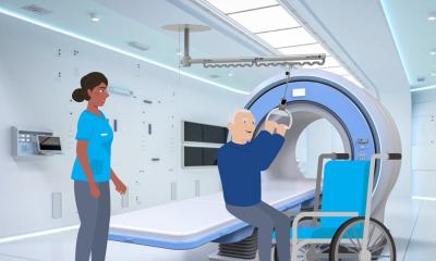
Introducing the session, Dr Simone Busoni, of the ESR Radiology MR Safety and Quality Subcommittee, was clear that the MRI environment is complex and that the consequences of getting it wrong are serious – ranging from tissue heating to equipment damage from projectile effects.1 A recent ESR survey2 underlined the scale of the challenge: In approximately 12% of responding hospitals, more than one implant-related accident had occurred in the preceding five years, and almost two thirds reported that they were not aware of existing MR safety guidelines. Furthermore, only 60-70% of patients with cardiac conditional devices were actually being scanned, despite it being safe and appropriate to do so. ‘We cannot overestimate the risk, because this may result in patients being excluded from the benefits of an MRI examination, but also, we cannot underestimate the risk, which of course may lead to accidents and incidents,’ Busoni outlined the dilemma.
Safety walkthrough – from bore entry to active sequence
Prof. Siegfried Trattnig, Professor of Radiology at the Medical University of Vienna, pointed out the increasing frequency of patients with implants. ‘This is a really relevant topic,’ he noted. The expert walked his audience through the risk scenarios associated with the different phases of the MRI exam: before the patient enters the bore, inside the MRI scanner, and during an active sequence. Each of these phases involves different physical forces – translational attraction, torque, radiofrequency (RF) heating, and gradient-induced vibration – and each must be considered separately.

The complexity of these effects becomes even more pronounced when implants are involved. While the “MR Safe” and “MR Unsafe” labels are relatively straightforward, “MR Conditional” – applied to devices that are safe only under specified circumstances – is the most common classification for passive implants such as aneurysm clips, stents, or hip prostheses – and also the most ambiguous. The conditions attached to these implants can include limits on field strength, spatial field gradient, energy deposition rates, and other factors. Following these conditions – which are defined by the implant manufacturer – is crucial for safe MR operation, Trattnig stressed.
Some implants carry conditions that are far from straightforward. Self-expanding stents, magnetic growing rods, and external fixation devices used in orthopaedic patients, for instance, come with highly specific requirements that are not always easy to verify or control. Vendor-provided field maps are an essential resource here, and software tools from Philips, GE Healthcare, and Siemens Healthineers can assist further by allowing manual entry of implant-specific parameters and flagging critical zones visually. For teams who regularly scan patients with implants, Trattnig offered a practical tip: ‘If you deal with a high volume of such patients, it is worth creating your own archive.’
Implant-related artifacts and how to handle them
Many implants are not only potential safety issues in MR imaging, but can also significantly impair image quality, Professor Edwin Oei, Professor of Musculoskeletal Imaging at Erasmus Medical Centre in Rotterdam, pointed out. This is especially relevant when the device itself is the source of pathology – for example, due to implant-related infection, pseudotumour formation, aseptic loosening or neural compression.3,4
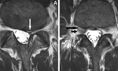
Depending on their material, size, position and geometric complexity, implants distort the MRI signal in different ways.5,6 Oei illustrated the three main effects:

Image source: Aboelmagd S, Malcolm P, Toms A, Reports in Medical Imaging 2014 (CC BY-NC 3.0)
- signal loss and signal pileup – presenting as overly dark or bright areas, respectively. In geometrically simple implants, this manifests as the so-called four-leaf clover effect;
- geometric distortion – a result of spatial signal encoding gone wrong; and
- failed fat suppression. ‘This can be problematic when we are looking for bone marrow oedema or soft tissue oedema, we may simply miss it’, Oei cautioned.
While the use of lower field strengths has been shown to mitigate these effects,7 the expert pointed out the limited availability of 0.55T scanners, leaving 1.5T MRI as the best practical compromise between signal-to-noise ratio and artefact reduction. Image quality can also be increased by adjusting MRI protocols – including sequence type, bandwidth, slice thickness, matrix size, and encoding direction.8 For the most demanding cases, advanced metal artefact reduction sequences, including 3D multi-spectral imaging techniques such as SEMAC, MAVRIC and O-MAR, can dramatically improve visualisation of tissues immediately adjacent to an implant.9
These sequences are now widely available and continue to improve, but they come with trade-offs, the expert noted: Scan times are longer, while overall image quality is decreased compared to regular sequences, higher tissue heating, and limited availability for some image weightings. In practice, Oei recommended combining them with standard sequences, which remain perfectly adequate for areas further from the implant.
From label to patient: the radiographer’s role
The final presentation brought the session’s themes into the day-to-day reality of clinical practice. Christos Tsiotsios, MSc, a radiographer with specialist expertise in MR safety, framed the challenge succinctly: ‘It’s not just about identifying an implant – it is about interpreting the conditions and maintaining workflow efficiency without compromising patient safety.’ For this, he urged his colleagues to pay close attention to the specific Do’s and Don’ts of any “MR Conditional” label: ‘An implant labelled for 1.5 Tesla is not automatically safe at a lower or higher magnetic field. Or, if a patient is positioned off-centre for a shoulder scan, the implant may experience a higher spatial gradient than the manufacturer’s specification.’ For high-risk passive implants, Tsiotsios advocated measuring RF exposure not through the specific absorption rate (SAR), but with the B1+rms metric, as it provides a consistent, patient-independent measure of the RF field actually delivered.
A previous MRI does not ensure patient safety in any way for future MRI examinations
Christos Tsiotsios
The less is known about an implant, the more cautious radiographers need to be, Tsiotsios advised, describing his approach as an “identification pyramid”: starting with the implant card and manufacturer’s website as the gold standard, then working down through electronic health records, surgical notes, specialist databases such as MRIsafety.com or MagResource.com, prior imaging, and finally plain radiography or CT. Crucially, he warned, ‘a previous MRI does not ensure patient safety in any way for future MRI examinations.’ The expert singled out intracranial aneurysm clips as a particular category of concern: scanning must not proceed until the specific manufacturer, model, and type have been confirmed, as the consequences of error can be fatal.10 ‘For unknown passive implants, the policy should be a collaborative effort between the supervising physician, the radiographer, and the medical physicist,’ Tsiotsios advised.
Looking ahead, the expert discussed the potential of artificial intelligence to assist with pre-examination implant identification by analysing electronic health records and prior imaging, with studies already reporting accuracy above 80%.11 While these AI system show great promise for enhancing workflow efficiency and MRI safety, data privacy and validation challenges must be addressed, and human oversight remains essential, he concluded: ‘The label provides the limits, but the clinical team ensures patient safety.’
Profiles:
Dr Simone Busoni is a senior medical physicist at the UOC Medical Physics department of Careggi University Hospital in Florence, Italy, where he holds a Level III Radiation Protection Expert qualification — the highest certification level in Italy. He serves as coordinator of the EFOMP working group responsible for revising Policy Statement 14 on MRI Safety, the European reference framework for safe MRI practice, and is a member of the ESR Radiology MR Safety and Quality Subcommittee.
Prof. Siegfried Trattnig is Professor of Radiology with a specialist focus on high-field MRI at the Medical University of Vienna, Austria. Since 2000, he has served as Medical Director of the University of Vienna's high-field MR research scanner, and since its founding in 2003, as Director of the High Field MR Centre of Excellence at the Medical University of Vienna. He has been a member of more than 50 committees across the major international societies in radiology, orthopaedics, and MR imaging, including the ESR Research Committee Board; he has served as Chairman of the European Imaging Biomarker Alliance of the ESR, Chairman of the ISMRM Musculoskeletal Study Group, and Director of the School of MRI of the ESMRMB. He sits on the editorial boards of seven peer-reviewed journals and has authored over 500 indexed publications in PubMed as well as 24 book chapters.
Edwin H.G. Oei, MD, PhD, is Associate Professor of Musculoskeletal Imaging and Section Chief of Musculoskeletal Radiology in the Department of Radiology and Nuclear Medicine at Erasmus MC, University Medical Center, Rotterdam, the Netherlands. As principal investigator of the Advanced Musculoskeletal Imaging Research group at Erasmus MC (ADMIRE), his research focuses on multi-parametric MRI, CT, and ultrasound imaging of diseased joints. His work encompasses the technical development, validation, and implementation of novel MR techniques in large clinical and population-based studies.
Christos Tsiotsios, MSc, is Head of Radiographers at Markides MRI and Diagnostic Imaging in Limassol, Cyprus, and a part-time instructor at the European University of Cyprus in Nicosia. He specialises in MR safety, with a particular focus on the practical management of patients with passive implants in clinical radiography practice.
References:
- Nordin LE, Åberg K, Kihlborg J et al.: ESR Essentials: basic physics of MR safety—practice recommendations by the European Society for Magnetic Resonance in Medicine and Biology; European Radiology 2024
- European Society of Radiology (ESR): The European MR safety landscape; Insights into Imaging 2024
- Pogliacomi F, Schiavi P, Calderazzi F et al.: Is there a relation between clinical scores and serum ion levels after MoM-THA? One year results in 383 implants; Acta Biomedica 2020
- Bruschetta A, Palco M, Fenga D et al.: How to Manage Metallosis: A Retrospective Cohort Analysis after Revision Hip Surgery; Journal of Clinical Medicine 2023
- Feuerriegel GC, Sutter R: Managing hardware-related metal artifacts in MRI: current and evolving techniques; Skeletal Radiology 2024
- Kim Yj, Yoon D, Song SO, Lee S, Choo HJ, Ryu J: Metal Artifacts in Postoperative Spine MRI: Strategies and Limitations; Investigative Magnetic Resonance Imaging 2025
- Luitjens J, Ziegeler K, Yoon D et al.: Improved metal suppression using new generation low-field MRI: a biophantom feasibility study; Skeletal Radiology 2024
- Aboelmagd S, Malcolm P, Toms A: Magnetic resonance imaging of metal artifact reduction sequences in the assessment of metal-on-metal hip prostheses; Reports in Medical Imaging 2014
- Ziegeler K, Yoon D, Hoff M, Theologis AA: Metal Suppression Magnetic Resonance Imaging Techniques in Orthopaedic and Spine Surgery; Journal of the American Academy of Orthopedic Surgeons 2025
- Klucznik RP, Carrier DA, Pyka R, Haid RW: Placement of a ferromagnetic intracerebral aneurysm clip in a magnetic field with a fatal outcome; Radiology 1993
- Valtchinov VI, Lacson R, Wang AW, Khorasani R: Comparing Artificial Intelligence Approaches to Retrieve Clinical Reports Documenting Implantable Devices Posing MRI Safety Risks; Journal of the American College of Radiology 2020
19.08.2026