Tumor ablation brings promise
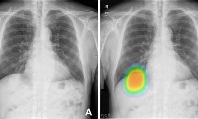
More and more patients with lung cancer are being treated with thermo-ablation techniques, and radiologists are increasingly being confronted with images from these patients that may be difficult to interpret.

“Follow-up is very challenging, and if for no other reason, this presentation will be of interest to many attendees at ESTI to understand the conditions that may manifest after treatment by ablation,” said Benoît Ghaye, MD. “Where there are 20 years of experience with thermo-ablation for hepatic cancers, the application of the technique to pulmonary tumors was introduced only 10 years ago, and then in a restrained manner,” he said. Recently, however, the use of these techniques has seen a significant take-up in the clinic. The larger size of the needles for radio-frequency ablation (RFA) of liver tumors was the primary barrier to adoption for pulmonary applications.
Adapting needles to the lung reduced this challenge, but a risk of complications continued to delay wider use. The concern was the potential for gas generated by the thermal ablation to provoke cerebral embolizations. “Applying heat to a tumor is not unlike cooking a steak,” said Ghaye; “there is smoke, or gas. Applying this energy in the liver, the gas passes by the venous system until it comes to the pulmonary capillaries, and the body does not generally have a problem.” “On the other hand, when we heat lesions in the lungs, unfortunately the return of the gas is not by the right side of the heart, but on the left, and it passes directly into the systemic circulation,” he explained.
Subsequent experience showed thermoablation caused fewer problems than feared at the beginning, such that the complications to the circulatory system remain theoretical, rare in real experience. Meanwhile, the enhancement to the technology allowed a more rapid application of heat with lower gas emission. With RFA there remains a complication with the carbonization of the tissue from the RF needles that accumulates on the needle and tends to insulate the energy transfer.
“There are techniques for reducing this effect, Ghaye said, and technical improvements include RFA needles cooled by water at four degrees Celsius that cool down the tissue that is in contact with the needle.” Using these tools and techniques allows the clinician to treat a greater volume, but RFA is not an ideal treatment of all types of lung lesions. In the past five years microwave ablation (MWA) has been increasingly used for ablation of pulmonary tumors. “When I first used this approach in 2007, it was like using a bazooka to swat a fly,” said Ghaye, as microwave can obtain higher intratumoral temperature than RFA.
“In early applications, the energy level was much too high, and at congresses we saw images of complications from microwave ablation with large zones of necroses and cavitations. The algorithms for the application of energy have been since greatly improved, and there are few complications,” he said. “Microwave ablation seems to be a technique better adapted than RF for treating some types of pulmonary tumors,” he said. The risk of carbonization was significantly reduced, for example.
Another advantage, Ghaye explained, is a reduced loss of heat through large blood vessels in contact with the tumor.” These vessels tend to absorb heat in what is called the “heat-sink effect”, making it impossible to treat the part of the tumor that is in contact with these vessels. As a result, there is a risk of recurrence for the cancer from these portions of the tumor left untreated. This risk is reduced due to the uniform and controlled application of heat with MWA. “We can actually have a higher temperature with microwave without the risk of carbonization of tissue,” said Ghaye. “Cystic lesions are much easier to treat; there are less interferences with pacemakers, and where we need patient grounding pads and a risk of burns using RF, this is not required for MWA.”
“In my experience, it is simpler and quicker to treat with microwave ablation, but it is more expensive,” he said. With RFA needles costing one third to half the price of MWA, the newer procedure is typically only performed in major university centers that can absorb the cost. This issue varies from one country to another, he said. “In Belgium we have the problem that neither the needles for RFA nor for MWA are reimbursed by the social insurance for pulmonary interventions.”“So while we may prefer to use MWA for most patients, RFA remains more affordable,” he said. New evidence supporting MWA is expected to be published in the near future, and six manufacturers are introducing new systems. During his presentation, Ghaye said he will also discuss other technologies being applied for ablating pulmonary tumors.
“Cryo-ablation has mainly been used in the kidney, but bringing it into the lung shows bleeding complications,” he said. “However, cryo-ablation has other advantages, in particular for ablation of large lesions in contact with or invading the chest wall.” “There are also treatments by laser, and finally there is a completely new technique called “irreversible electroporation”. To date there is only one paper that has been published, and this was for animal studies. We see the potential for great advantages in man, because there is not the heat-sink effect as with RFA and to an extent with MWA.
For your diary
Pulmonary RFA and MWA –
current status and trends
Session “Thoracic Intervention”
Benoit Ghaye, Brussels, Belgium
Sunday June 24 at 11:20 – 11:50
####
Profile
Benoît Ghaye has served as the head of Cardio-Thoracic Imaging Section, Department of Medical Imaging, University of Liège in Belgium. A founding member of the European Society of Thoracic Imaging (ESTI), he is currently the chairman for the Electronic Media Committee and also serves as Chairman of subcommittee on Chest for the European Society of Radiology (ESR). The author or co-author of more than 75 scientific papers, he has contributed numerous chapters to books, an is the leading author of the text on percutaneous image-guided interventional procedures in the thorax.
25.06.2012