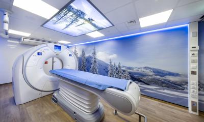
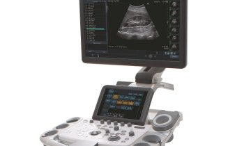
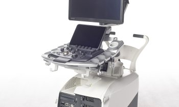
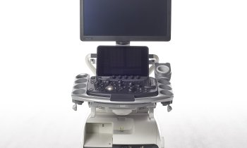
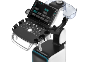
Fusion and Fly Thru - the new Aplio 500
Catastrophes draw people closer, as demonstrated by the development of the new high-end ultrasound scanner Aplio 500 from Toshiba. The clinical evaluation period took place during the tsunami and the nuclear catastrophe in Fukusima. Professor Thomas Fischer at the Radiological Institute, Charité Clinic in Berlin, was impressed by the enormous commitment shown by the Japanese firm’s engineers in the development and optimisation of this equipment during the clinical test period.
‘Events in Fukushima torpedoed the entire development effort,’ he explained. ‘On the one hand, this was because certain kinds of work could not be carried out in Japan and, on the other, because some of the developers were personally affected by the catastrophe. To honour the tireless efforts of these people we put even more efforts and energy into the clinical evaluation of the technology. Despite the adverse conditions, our requests and suggestions were sometimes literally implemented overnight. Step by step, we jointly developed a product that now exceeds expectations by far.’
The fact that Toshiba handed over the prototype of the new Aplio Series for clinical testing at a very early stage was a prerequisite for this success. Prof. Fischer: ‘We did not receive a completely finished product for testing purposes but could provide our input with regards to the requested settings and presets. I valued this because it enabled us to improve the image step by step, to optimise our working routines and to develop new ways of image processing. The combination of technicians on one side and clinicians on the other turned out to be a true recipe for success.’
Better images and more diagnostic facilities
The result of this mutual commitment exceeds the capabilities of its predecessor, the Aplio XG, with the quality of the B-image improved even further. The smart fusion tool for abdominal imaging -- a focus of the professor's work -- i.e. a new kind of technology for the fusion of ultrasound images with images produced in other modalities, is also new. ‘This feature,’ he explains, ‘allows me to routinely import CT data sets and to combine these live with ultrasound image data. So I can move through the CT and ultrasound image in real time, change details of the CT examination, directly compare results with one another, carry out punctures and apply ultrasound contrast media. The entire process is relatively easy and therefore very suitable for clinical routine.’
The procedure’s advantage for patients is obvious. Ultrasound is not always the first imaging procedure a patient has undergone -- many bring along the results of CT examinations for further clarification.
Previously, the difficulty was in finding and scanning the exact same location with ultrasound, particularly in case of liver segment VIII in subcapsular position. With the fusion and consolidation of images, that uncertainty is no longer an issue because the result can be marked on the CT image and the identical position can then be scanned with ultrasound so that intercostal sections can also be compared. ‘Additionally, I have a lot of freedom to optimise the examination,’ he explains. ‘For example, I can compare CT density differentiations with the B-image characteristics of a target lesion, or use contrast media for the same set of data, as the CT examination is often carried out in two phases as part of staging and is not optimised for the liver. This improves my diagnostic capabilities.’
The next step, he adds, would be to import a calculated set of CT data and then carry out a puncture or radio frequency ablation via ultrasound. ‘This means that both the ultrasound examiner and the nurse do not need to be exposed to the radiation levels caused by a CT scan, yet with comparable diagnostic certainty.’
Image fusion obviously also works with other modalities. However, a challenge in clinical routine (at least in Germany) could be that ultrasound is the responsibility of different specialists, such as internists, radiologists or gynaecologists, who then would also need the necessary knowledge and experience in CT or MRI imaging.
Volume and contrast imaging on a new level
A further highlight of the Aplio 500 is Fly Thru, a so far unique form of virtual imaging with ultrasound. ‘Using ultrasound, the images of the virtual journey through the intestine, for instance, are impressive and offer, among other things, the potential for endosonography.
‘However, this type of examination requires the highest specifications in terms of the equipment.’ Ideally it should be carried out with a 4-D probe that can be used with contrast media and allows a high volume rate.
A further option would be a balloon in the delay path, which fits around the tumour so that the surface can be detected even more accurately,’ the professor explains. ‘What further uses Fly Thru may have will -- as with all completely new procedures – will be seen in the coming months; further applications could be in gall ducts or milk ducts imaging. In principle, all fluid-filled cavities are virtually accessible in this way.
The advantages of real-time elastography, which clearly optimises the examination of the abdomen and mammary gland and further small-part applications, have already been ascertained. Differences in tissue density can also be detected and quantified for ageing, tumour characterisation and infectious changes in the tissue. The spectrum ranges from the liver to the prostate and breast ultrasound.
Improved user friendliness for contrast media administration is of particular added value for Dr Fischer: ‘Contrast enhanced ultrasound these days is very much a routine procedure, especially in abdominal diagnostics. With the Aplio 500 its application has been even further simplified. The procedure is becoming increasingly important, such as in the case of patients with kidney problems where neither CT nor MRI scanning is possible. Ultrasound offers the only diagnostic alternative here.
‘And.’ Prof. Fischer adds, ‘through simplified use via presets, even less experienced examiners can achieve accurate results. Moreover, the procedure is so robust that the amount of contrast medium can be lowered to 1.2ml without any loss in image quality.’ The import of raw data, quantification of these data and optimised imaging of contrast enrichment have been further simplified.
However, there is one problem with these new technologies: Lack of time and staff in clinical routine to learn about the innovations and therefore to introduce them comprehensively into clinical routine. ‘Due to financial pressures, diagnostics frequently lags behind the technological capabilities,’ Prof. Fischer concludes. There is a need for clear training concepts and payment for highly specialised applications.
"State of the Art in Ultrasound Imaging - New Tools and Perspectives", Toshiba Lunch Symposium, Sunday, August 28, 12:15-13:45, Hall F1
Thomas Fischer
Since 2007, Professor Thomas Fischer has directed ultrasound diagnostics at the Radiology Institute at the Charité Clinic in Berlin, and, from 2009, has also headed the ultrasound research laboratory at the Charité, which specialises in testing new ultrasound procedures and techniques in complementary studies.
He is also a member of the European Society of Radiology (ESR), the German Radiology Society (DRG), the German Society for Ultrasound in Medicine (DEGUM) and an honorary member of the Polish Society of Ultrasound (PUS).
Last year Prof. Fischer received the Scientific Presentation Award for the best paper at the European Congress of Radiology.
In 2011, received the Herbert M Stauffer Prize for his scientific paper Detection of Rheumatoid Arthritis Using Non-Specific Contrast Enhanced Fluorescence, classed as the best basic science paper.
25.08.2011
- breast cancer (544)
- elastography (89)
- imaging (1487)
- medical technology (1447)
- obstetrics (121)
- ultrasound (721)