Ultrasound-MRI fusion imaging
Promising research aims for precise, consistent, gentler prostate biopsies
Tissue removal is currently the only way to verify suspicious lesions in the prostate. It is also a key requirement before treating prostate carcinomas. The standard biopsy is performed with transrectal ultrasound (TRUS) to guide the needle in the right direction.
However, the ultrasound visualises only general outlines of the prostate and cannot reliably detect the location of the carcinoma. This means that tissue samples are, in effect, taken blindly and randomly in sextant regions of the prostate, leading to varying success rates and often to an increasing number of biopsies.
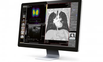
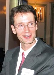
Philips Research is working on an ultrasound-MRI image fusion system to achieve greater accuracy during biopsies. The new technique superimposes pre-captured MRI scans onto 2-D ultrasound images so that urologists can see both the biopsy needle and potential tumours simultaneously. The technology is currently being clinically evaluated at Radiology and Imaging Sciences Department, at the USA’s National Institutes of Health Clinical Centre (NIHCC), where physicist Dr Jochen Kruecker, Principal Member of Research Staff with Philips Research, is currently based. Karoline Laarmann (European Hospital) asked him how fusion-guided prostate biopsy works and about its future potential.
‘We already successfully perform fusion-guided biopsy for the kidney and liver,’ he explained. ‘The difference is that we use pre-procedural CT scanning in these applications. By contrast, MRI enables precise soft-tissue visualisation in the prostate. Up to now, the difficulty was to hit the MRI findings later on with the ultrasound. So the idea was to combine the best of both worlds: the accurate diagnostic information from pre-acquired MRI and the real-time anatomical information from the ultrasound image. This is done using an experimental multi-modality interventional workstation together with an electromagnetic tracking system. There are miniaturised sensor coils integrated in the biopsy needle, biopsy guide or ultrasound probe, which work like GPS chips. They receive electromagnetic signals that allow the system to determine the actual position of the devices. Then the system generates a fusion view of the live ultrasound image with the pre-acquired MR image and corrects it if necessary, for example, when a patient moves during the procedure.’
In the clinical studies, the primary target group will be patients who had a prior failed biopsy, Dr Kruecker explained. ‘In about 20% of cases, the standard sextant biopsy misses the cancer. So there is quite a high number of patients who have reason to believe that they have cancer because of other diagnostic tests, but as long as the biopsy does not supply evidence, therapy cannot be initiated. So the biopsy has to be re-done – and it’s not guaranteed that if you repeat a standard biopsy it will find the cancer that was missed the first time, especially when the cancer lesion is small, or located in a difficult area. Certainly, using the fusion-guided procedure may not improve results for all patients. MRI is not the perfect solution for everyone – it might miss certain cancers, or show something suspicious that turns out to be benign. So there will definitely have to be a selection depending on the specific diagnostic picture. Nevertheless, our first results show a high correlation between the suspicion level assigned by radiologists reading the MRI and our positive fusion-guided biopsy rate.’
The research is currently at the single-centre clinical study phase. ‘That means, we are doing both: the standard 12-core sextant biopsy with 2-D ultrasound imaging and then the targeted fusion-guided procedure,’ he explained. ‘The patient population includes men with elevated PSA and/or abnormal digital rectal examination; men with a history of elevated PSA but one or multiple negative TRUS-guided biopsies, and men with known low-grade prostate cancer who are on “watchful waiting”. Next, we are investigating the possibilities for a multicentre study, for example in the USA. Based on the clinical results from such a study, the decision can be made to start the development of a commercialised version of the tracking software. A preview of the results of the technology will be presented at the 2009 Annual Meeting of the American Urological Association (25-30 April).
Could this technique be applied in other fields? ‘We are demonstrating the feasibility of the technique now for prostate biopsy but it may have an even bigger impact on minimally invasive focal therapy like ablation procedures. In recent years, the idea of using focal therapy in the prostate has gained a lot of momentum, but the image guidance has always been the problem. Our technology may open the door to an effective delivery of focal therapy to the cancer lesions that are identified in the MRI. Therefore, at the NIHCC, we are just preparing to begin preclinical and clinical work to translate the technology from a diagnostic application to therapy.
01.05.2009