Article • Old technique & new technology
Optoacoustics: the sound of cells
For centuries, hands, eyes and ears were the physicians’ most important instruments when it came to detecting and diagnosing disease. Today, one of the traditional techniques, percussion, is being revived, supported by state-of-the-art technology and dressed in a new name: optoacoustics.
Report: Anja Behringer
In one of the most exciting visionary ideas in modern healthcare short laser pulses (optics) are transmitted to tissue where they generate ultrasound signals (acoustics) that allow the identification of cells and diseases in the body. The advantages of this technology? No ionised radiation, no invasive procedure.
Pioneer of clinical optoacoustics is Professor Vasilis Ntziachristos, Chair for Biological Imaging at the Technical University Munich (TUM), Germany, and Director of the Institute of Biological and Medical Imaging (IBMI) at Helmholtz Zentrum, Munich. His groundbreaking research not only sparks hope for cancer patients but also opens new diagnostic perspectives for Alzheimer’s, diabetes and dermatological diseases.
Multi-spectral optoacoustic tomography (MSOT)
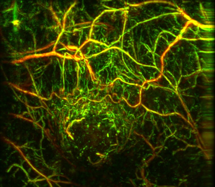
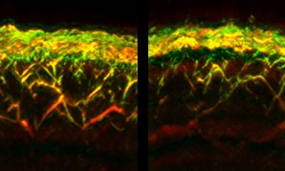
The laser pulses penetrate the body where they are absorbed differently, depending on their wavelength and on the type of target tissue. These laser pulses create a minute rise in temperature which expands the tissue. Those equally minute movements generate acoustic signals – with each type of tissue producing unique signals. A blood cell for example “sounds” very different from a skin cell. Ultrasound detectors on the skin surface register these different signals and a computer generates the corresponding 3-D image. Thus, single cells, for example cancer cells, can be detected. This is a major advantage compared to ultrasound, which cannot differentiate on this level, Ntziachristos explains. Currently, multi-spectral optoacoustic tomography shows its potential particularly well in aggressive melanoma cells. But their unique sound also gives away other cell types, which might allow surgeons to check accurately during a tumour resection whether indeed all cancer cells were removed.
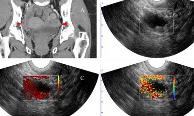
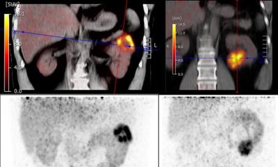
Following successful animal studies the procedure is now being tested in human volunteers. Different clinical studies are currently being conducted for breast and thyroid cancer and peripheral atherosclerosis. To display the images, another expert in medical imaging, Professor Dr Daniel Razansky of Helmholtz Zentrum Munich, is developing an affordable diagnostic device for clinical use in the operating room (OR). While the device today costs around €200,000, Ntziachristos considers a future price tag of €50 to be realistic. Thus, in 2011 he and two partners founded the spin-off iThera Medical in Munich to fine-tune the product for a market launch. This requires capital.
Awards for visionary research
When it comes to raising capital, the many awards Ntziachristos, a qualified electrical engineer, has collected over the past few years obviously help to give investors peace of mind. In 2013, he received the Leibniz Prize of the German Research Foundation (DFG) and last year he was awarded – for the second time – the ERC Advanced Grant of the European Research Council. That grant of €2.49 million will be disbursed over a period of five years. The funds will be used to develop a portable device for human patients. As to market maturity the Helmholtz Zentrum did not provide any information since the product is still under development.
While working on the marketability of the device, Ntziachristos’ research is also addressing the major limitations of optoacoustics: the laser cannot penetrate deeper than four to five centimetres and the sound loses quality the deeper it moves into the tissue. Thus, liver studies, for example, are impossible.
Ntziachristos’s colleague Razansky is particularly interested in the molecular level, the biochemical reaction in the cell. Whatever the individual research interests of the two scientists are, the overarching aim of the team is early detection of diseases and tracking of disease progression so as to save time-to-diagnosis and money and above all to help patients by improving prognosis. Additionally, the optoacoustic imaging pioneers think even further. They envisage new interdisciplinary fields of medical science and new areas of application, such as the diagnosis of inflammation or metabolic disorders and neurology. To test their ideas the researchers successfully applied for Horizon 2020, the major European Union research and innovation programme for 2014 to 2020.
Profile:
Research carried out by Professor Vasilis Ntziachristos encompasses the development of new methods and devices for biological and medical imaging, focusing on innovative non-invasive approaches that visualise previously unseen physiological and molecular processes in tissues. His research also aims to translate these methods to advance biological discovery, accelerate drug development and offer efficient methods for diagnostics and theranostics. Ntziachristos studied electrical engineering at Aristotle University, Thessaloniki, Greece, and gained Master’s and doctoral degrees from the Bioengineering Department at the University of Pennsylvania, USA. He was assistant professor and director of the Laboratory for Bio-Optics and Molecular Imaging at Harvard University and Massachusetts General Hospital. He now directs the Biological and Medical Imaging Institute at Helmholtz Zentrum Munich, Germany.
29.01.2018