Article • New scan
Hyperpolarised MRI detects inflammation after heart attack
A new type of scan that can detect cardiac inflammation may help tailor treatment for patients who have suffered a heart attack, according to research findings presented at the British Cardiovascular Society (BCS) conference in June.
Report: Mark Nicholls
Developed in the UK, hyperpolarised MRI (h-MRI) enables cardiologists to see more detail of healthy and diseased hearts than conventional scans, including the level of inflammation post-myocardial infarction.
Detecting inflammation in the heart
Funded by the British Heart Foundation (BHF) the University of Oxford researchers now believe h-MRI could help scientists develop and monitor the effects of new, inflammation-targeting treatments that may improve the heart’s healing process in heart attack patients. The discovery could have significant benefits because the hearts of patients who have suffered a myocardial infarction (MI) can undergo continued inflammation even during the healing process, and despite receiving emergency treatment.
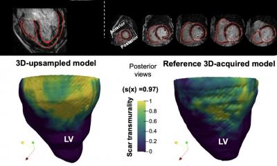
Yet research into why this may happen remains limited as conventional scans cannot detect or measure inflammation in the heart, whereas h-MRI can. Conventional MRI measures how protons change position when exposed to a magnetic field, but h-MRI works by producing images from carbon molecules, which make up the energy sources needed to help the heart pump and can offer doctors a clearer picture of inflammation in the heart.
h-MRI as biomarker and pharmacological target
For the study, the team measured production of lactate in the damaged heart tissue of rats and found that after a heart attack immune cells within the injured heart muscle become active and are reprogrammed to make lactate, leading to inflammation in the heart. By monitoring how much lactate is produced in the damaged tissue, they were able to identify and measure the level of inflammation in the heart. Following on from that, the team administered the experimental drug 2-deoxyglucose (2-DG) to the rats after the heart attack, to try and combat the inflammation. Using h-MRI to monitor the heart’s response, they found that 2-DG reduced lactate production and inflammation, and improved the heart’s function.
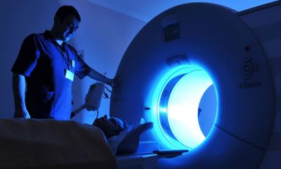
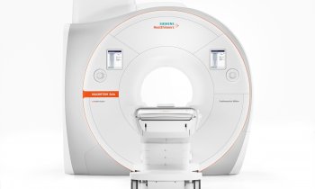
The cutting edge scanning system

They showed that high hyperpolarised lactate signal in the days after myocardial infarction is caused by macrophage-driven inflammation, and reflects not just the number of inflammatory cells infiltrating the myocardium but also the inflammatory phenotype of those cells.
The Oxford scientists – one of the first groups to use h-MRI to study the human heart – believe that h-MRI will not only detect inflammation in damaged heart tissue but also could provide a novel method for the detection of myocardial inflammation with high translational potential as both a biomarker and novel potential pharmacological target. ‘H-MRI is a cutting-edge scanning system which allows doctors to better understand the innermost workings of the heart non-invasively, and how they become abnormal in heart disease,’ Dr Andrew Lewis, Specialist Registrar in Cardiology at the Great Western Hospital in Swindon, summed up. ‘In this research, we used h-MRI imaging to capture the healing processes in experimental models after a heart attack, and also tested new treatments to improve the heart’s recovery.
‘Our work has identified several forms of heart disease where this technique could be used to improve diagnosis and treatment,’ Lewis added. This is incredibly exciting, and we intend to move forward with patient studies as quickly as possible.’
Profile:
Andrew Lewis MD is a Specialist Registrar in Cardiology at the Great Western Hospital in Swindon, UK, and won the Young Investigator’s Prize Competition at the British Cardiovascular Society conference in Manchester for his research into myocardial inflammation imaging.
30.08.2017