News • Microscopy
Dense vessel network regulates formation of thrombocytes in bone marrow
Scientists from the University of Würzburg give fascinating 3D-insights into the bone marrow, and successfully elucidated new details about the process of thrombocyte generation.
These important findings could contribute to optimized therapeutic approaches for patients with bleeding disorders.
Thrombocytes, also known as platelets, have an important role in wound healing and tissue repair at sites of vascular damage by facilitating blood coagulation. As they possess only a short lifespan, new thrombocytes need to be constantly generated in the body. Platelets originate from the bone marrow where giant precursor cells, so-called megakaryocytes, undergo a complex maturation process and finally release platelets into the bloodstream.
The nursery of thrombocytes: calmer than one thought
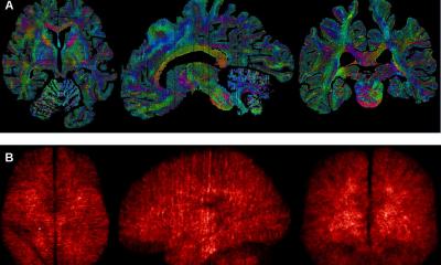
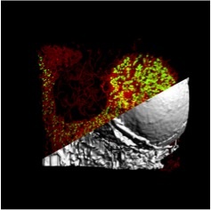
So far, these precursor cells have been believed to migrate through the bone marrow to reach the vessel. By applying a combination of modern microscopy methods and computer simulations, researchers from the Rudolf Virchow Center for Experimental Biomedicine and the University Hospital of Würzburg were now able to disprove this theory. The team of Prof. Katrin Heinze and Dr. David Stegner could show that most megakaryocytes already originate from the vascular niche, that is in close proximity to the vessel. The remaining precursor cells are uniformly distributed in the bone marrow and are typically so large and flexible in shape that they can reach the vessel by small (wobble) movements in the dense vessel network of the bone marrow.
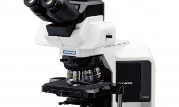
"Only the combination of all information from intravital and light-sheet microscopy has made these findings possible," says Professor Katrin Heinze, director of the study. She further explains: "In addition, we were able to show how the 3D images of vessels and cells become perfect biological templates for realistic, eye-opening simulations of cell distributions in the bone marrow".
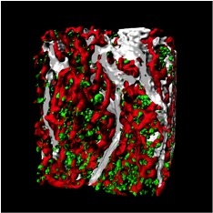
Complementary microscopy methods allow a virtual journey through the bone marrow
Dr. David Stegner, corresponding author of the new study, adds: "We have long suspected that the existing models of thrombopoiesis are insufficient or false, but it was difficult to prove this experimentally. This has now been achieved by high-resolution insights into the intact bone!“ The two leaders of the study point out that their collaboration was not only a stroke of luck, but a future-oriented decision that will continue for a long time to come.
"Based on our findings, new treatment strategies for diseases that are associated with reduced platelet formation could be developed in the future", hopes Dr. Stegner. The researchers from Würzburg recently published their new findings in the scientific journal Nature Communications.
Source: Rudolf-Virchow-Zentrum für Experimentelle Biomedizin der Universität Würzburg
27.07.2017