News • Neurosurgery
Microscope imaging system integrates virtual reality technology
Joshua Bederson, MD, Professor and System Chair for the Department of Neurosurgery at Mount Sinai Health System, is the first neurosurgeon to use CaptiView – a microscope image injection system from Leica Microsystems that overlays critical virtual reality imaging directly onto the brain when viewed through the eyepiece, known as the ocular, during surgery.
This new microscope technology allows images of chosen objects, including original CT, MRI and angiogram datasets, to be superimposed, or ‘injected,’ directly into the neurosurgeon’s eyepiece during microscopic surgery. “This next-generation augmented virtual reality tool provides real-time information in ways never before realized,” says Dr. Bederson, who is now using the technology for all of his cases. He worked closely with Leica Microsystems and Brainlab® to develop the surgical navigation tool.
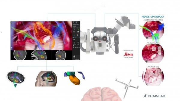
The CaptiView image injection system utilizes Brainlab® Cranial 3.1 Navigation Software in conjunction with a Leica M530 OH6 microscope. The heads-up display provides neurovascular and fiber-track information in 2D or 3D as well as on-screen video overlays visible through the ocular. The microscope integration also allows the surgeon to switch views in the eyepiece, toggling between live and pre-operative anatomical images using handle control buttons or footswitch for ease of use and uninterrupted workflow. Markers attached to the microscope enable positional tracking and autofocus.
This new technology will be utilized alongside Surgical Navigation Advanced Platform (SNAP) developed by Surgical Theater, LLC, which is a standard feature in the operating room. SNAP provides advanced 3D visualization technology that gives surgeons an intraoperative and patient-specific 3D environment to plan and understand surgical approaches. “We are driving and advancing the development of next-generation simulation and virtual reality technology, which can help improve patient outcomes and solve neurosurgical challenges,” says Dr. Bederson.
Source: Mount Sinai Health System
18.08.2016