Article • Hybrid OR
Thumbs up for new C-arm system
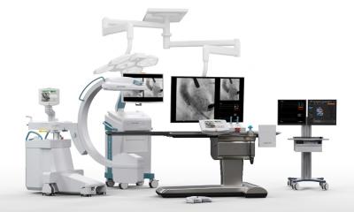
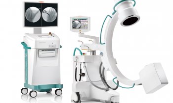
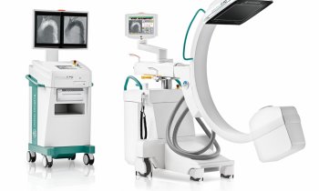
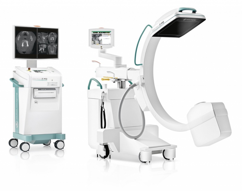
3D imaging is continuously improving, with devices simultaneously becoming more manageable and mobile. The new C-arm system Ziehm Vision RFD 3D is opening up a new dimension. The device was tested by Dr Jan-Sven Jarvers, orthopaedic and trauma surgery specialist at the University Hospital Leipzig, and was introduced last September during the Eurospine Congress in Copenhagen. ‘In the future, newly-developed intraoperative 3D imaging may replace postoperative CT scans and reduce the number of repeated surgical interventions significantly,’ he concludes.
Report: Marcel Rasch
Outstanding image quality and large volume
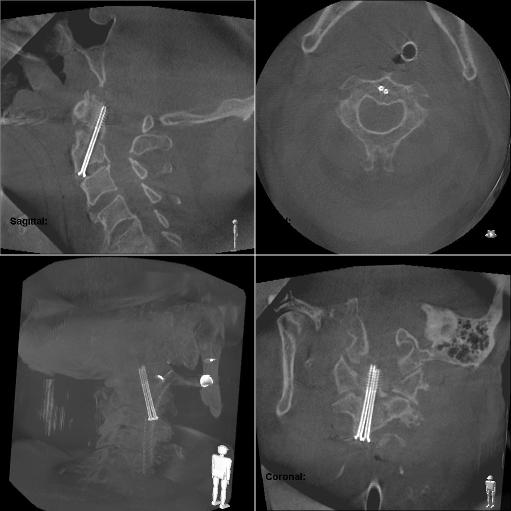
‘A big advantage,’ he adds, ‘is the outstanding image quality which, unlike its predecessors, facilitates a larger volume of the areas to be visualised.’ With an edge length of 16cm in the scan volume, up to seven cervical vertebrae can be visualised in one scan. ‘The system is of particular interest for imaging of the spine, because the area to be operated on can be assessed more clearly. Surgeons can monitor intraoperatively how the screws are positioned during fracture surgery and can change the screws if necessary or, respectively, carry on operating when the position is optimal.’
Previous image enhancers have two dimensions and miss the spine’s axial view. ‘This made it more difficult to assess the exact relation of the screws to the spinal canal,’ Jarvers observed. Furthermore, navigated interventions mostly use CT data sets as templates. Although this facilitates a 3-D view, it means having to refer back to images taken a few days before, where the patient may also have been lying in a different position. ‘Thanks to the 3-D C-arm, the patient’s current situation can be assessed during navigated surgery,’ Jarvers points out.
Fewer postoperative CTs and repeated interventions
Normally a hospital will carry out CT scans of the spine after surgery. However, not all hospitals adhere to this practice and some only perform postoperative CT scans in cases where the postoperative X-ray cannot be properly assessed, or if the patient develops unexpected symptoms or complications. This includes neurological symptoms such as numbness or paralysis, or increasing pain that cannot be explained. In such cases one must assume that screws are malpositioned, which could only be rectified through repeat surgery.
The intraoperative use of the three dimensional C-arm makes it possible to assess the placement of screws during surgery, lowering the risk of the need for a repeat intervention considerably.
The system also helps to assess whether the articular surface is well positioned during surgery for joint fractures. This makes the development of patients’ postoperative symptoms less likely. ‘You must also consider that postoperative CT scans always mean more radiation exposure and, in the case of repeated surgical interventions, also more anaesthetic administration,’ he warns. Also, time and cost for the hospital cannot be underestimated. The new Ziehm Vision RFD 3-D means patient safety goes hand in hand with simplification of daily routines in the operating theatre.
Good operability and almost no limitations
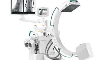
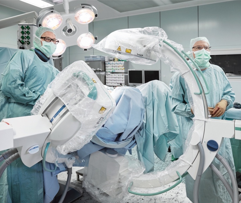
‘It is particularly convenient for nurses that everything is motorised,’ Jarvers says. The Ziehm Vision RFD 3-D can be motorised for all four movement axes, and automatically slows down when approaching a patient and automatically stops within a defined safety zone around the patient.
‘Fast processing is a further advantage,’ Jarvers adds. ‘Everything is very user friendly and geared towards working fast. Thanks to the large diameter of the C-arm we’ve had no problems with scanning, even with extremely obese patients.’
However, there can be limitations regarding image quality in patients with many endoprostheses. If surgery is carried out in the area where the cervical spine meets the thoracic spine in a patient with bilateral shoulder prostheses, those prostheses can limit image quality somewhat. ‘But,’ Jarvers adds, ‘the images are still good enough for us to safely assess the position of the screws. The software also helps with this because it automatically blends out many of the artefacts,’
One small detail should be noted. ‘The operating table should be made from carbon or at least have a carbon plate extension,’ the surgeon emphasis. ‘A table made from metal, or with metal on the sides, obviously makes resolution more difficult.’ Other than this, Jarvers is very convinced by the image quality: ‘I hope that intraoperative 3-D scans will become the standard.’
Profile:
Jan-Sven Jarvers MD gained his doctorate at the orthopaedics, trauma surgery and plastic surgery clinic, at Leipzig University Hospital, where he is now a specialist in orthopaedics and trauma surgery. His involvement with 3-D imaging and 3-D navigation began in 2007. His particular focus lies on spinal surgery.
03.08.2016