Obstetrics
New atlas will redefine OB/GYN imaging
Using the Arietta V70 from Hitachi, a French diagnostic imaging team is rewriting the book on obstetrics and gynaecology. Entitled the ‘Atlas d’échographie de fusion en gynécologie obstétrique’, the new edition by Jean-Marc Levaillant MD, and colleagues from the diagnostic imaging centres at the Bicêtre and Créteil hospitals in Paris, will be published before the end of 2015.
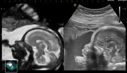
Driving the excitement behind the new atlas are unique double views of anatomy acquired using the Real-Time Virtual Sonography (RVS) feature on the Hitachi Aloka Arietta V70 ultrasound system, according to Laurence Gitz MD at the Prenatal Diagnostic Centre at Bicêtre. ‘Here we have a synchronisation of images rather than superimposing one image on top of the other, as with the usual fusion imaging. This means that we have a dynamic comparison with side-by-side viewing, which allows us to focus more clearly on a zone of interest and scrutinise better the anomaly,’ Gitz explained during a presentation at the French Radiology Congress.
The new atlas will include what she described enthusiastically as side-by-side views of a foetal cranium where the Hitachi echo images confirm a suspicion raised on the MRI images.
During the workshop, Naïma Chaibi MD noted that there could be uncertainties and disagreements about a diagnosis among clinicians when the MRI and ultrasound images are viewed separately. ‘In such cases where the condition is not clear on one modality, there is an advantage to bringing them together, in having both examinations simultaneously displayed,’ she said.
Chaibi currently leads a project to create a scoring method that will relate visualisations acquired by the Arietta V70 using RVS with an evaluation of risks for a patient. Originally developed for interventional radiology applications, Hitachi Aloka combined this new approach to fusion imaging with the company’s deep experience in OB/GYN examinations to develop the new application for the Arietta V70. After a scanner sequence is loaded onto the platform, a clinician clips a mini-sensor to the ultrasound probe that is tracked in a magnetic field projected from an antenna. According to Senior Product Manager Fréderic Philippe, the registration of the patient’s anatomy with the scanned data set is so robust that it can maintain the accuracy of a synchronised ultrasound exam even where there has been a shift of organs, or a foetus has moved.
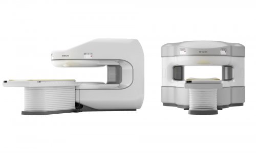
The lightweight and ergonomic design of the Arietta platform has been accelerated for the top-of-the-line V70 with the addition of high performance processing power that Philippe said is more than three-times more powerful than earlier versions.
The clinical advances Hitachi Aloka brings are the result of maintaining a horizontal, global approach to development that crosses ultrasound specialties, he added. ‘We bring to OB/GYN advanced tools and technologies originally developed in other clinical specialties. For example, the speckle tracking developed for cardiology has been applied to obstetrics for tracking a foetal heart. In bringing fusion developed for the liver and kidneys from radiology applications, we were able to show for the first time the foetal brain and have since shown the clinical relevance of echo fusion exams for diagnosing placenta abnormalities. Studies are now exploring cervical and ovarian cancers,’ he said.
‘We have stayed focused on a fundamentally different approach oriented to improving the clinical value of our technology. Sometimes ultrasound is used for clever marketing techniques, such as showing a snapshot of the unborn baby’s face. But this does not have a lot of clinical value. Our goal is to precisely monitor the development of the foetus or organ and detecting any abnormalities as early as possible,’ he said.
‘As a result of this commitment, starting in the first trimester of pregnancy, a clinician can see a level of information with an image quality that has never been available before. Clinicians have told us they are now able to see specific conditions of a foetus several weeks earlier in its development by using our probes and imaging platforms.’
16.11.2015