MS patients
Linköping University Hospital installs SyMRI NEURO
Linköping University Hospital in Sweden installs SyMRI NEURO from SyntheticMR in order to improve the follow-up of patients with Multiple Sclerosis (MS). SyMRI enables objective follow-up of brain atrophy through automatic calculation of Brain Parenchymal Fraction (BPF). After an initial pilot project, the aim is to take SyMRI into clinical use in Region Östergötland during 2016.

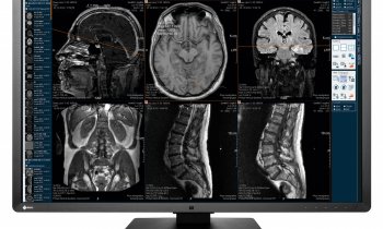
Magnetic resonance imaging (MRI) is especially good at visualizing soft tissue in the body and is used in the investigation and monitoring of MS. With current methods, however, physicians are for the most part forced to make subjective judgments, or visual estimations, of the images, which can make it difficult to obtain a good and comparable measure of disease development. Patients with MS can develop brain atrophy, or a loss of brain tissue. SyMRI NEURO generates a quantitative measure of brain atrophy by automatic calculation of BPF, thus enabling objective monitoring of disease development. Linköping University Hospital is now installing SyMRI to improve the monitoring of MS patients. The hospital will initially conduct a pilot project with the aim of taking SyMRI into clinical use in Region Östergötland during 2016.
“With SyMRI we can get an automatic measurement of brain atrophy, which means that we can now quantify the neurodegeneration that occurs in connection with MS. This allows for a better follow-up for our patients”, says Patrik Fägerstam, Neuroradiologist at Linköping University Hospital.
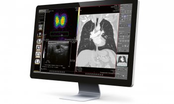
SyMRI NEURO will be installed in the hospital’s PACS from Sectra.
Source: SyntheticMR AB
28.08.2015