Art meets science
The future of endoscopy and imaging procedures
The future will be aesthetic or, put another way, Art meets Science. With this motto, the 43rd Congress of the German Society for Endoscopy and Imaging Procedures e.V., jointly held in Munich with six other specialist associations, demonstrated that aesthetic means the brilliance of images generated by the latest generation of X-ray, CT, MRI and ultrasound equipment.
Whilst those images have so far been of high-resolution, but mostly black and white, static and two-dimensional, this is increasingly changing. Visualisation from inside the body with panoramic functions, 3-D reconstruction, video films, functional and molecular-genetic imaging, marker-guided procedures and, finally, the interaction of the different visualisation procedures, open up new insights into as yet unknown perspectives that sometimes can be reminiscent of modern art.
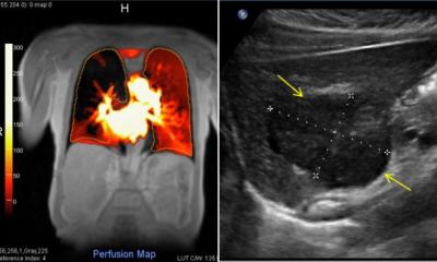
Christian Jennsen et al. write in their Thoughts on interfaces between Art and Science (Endoscopy Today 1/13): ‘With the help of high-resolution video-endoscopes it is now possible to visualise the finest mucosal structures of the embryo-genetically uniform aero-digestive tract in HD quality. Real and virtual colouring procedures highlight the finest structural characteristics of mucosal A few years ago the American forces succeeded in dramatically lowering the mortality of soldiers from gunshot wounds with the help of a new, haemostatic powder. These silicate crystals, which attach to a wound, not only stem external bleeding but also internal bleeding resulting from stomach or duodenal ulcers, tumours or rare types of vascular deformities. Named Hemospray, Cook Medical will introduce this new type of endosurfaces ingeniously. Confocal laser technology facilitates an insight into vital cell structures. Colour coded duplex and triplex ultrasound, as well as contrast-enhanced ultrasound procedures, show vessels and blood flow in organs and tumours in realtime. 3-D imaging allows fascinating insights into normal perfusion and tumour-associated neovascularisation.
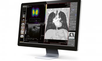
‘Multiplanary reconstructed and fused US, CT, SPECT and MRI images give a plastic impression of topographic relationships between organs, vascular structures and skeletal structures and their relation to pathological processes. This can, for instance turn them into threedimensional pointers for surgeons and interventionists for complicated procedures and also facilitates robotassociated surgical procedures and interventions.’
Colour is an essential contributor towards the current aesthetics in diagnosis because it adds to images another level of information. This in turn triggers emotions that are particularly well remembered in connection with what has been viewed. Guidelines on contrast-enhanced ultrasound procedures are now also available.
In the case of elastography, tissue elasticity levels are shown in colour. Harder tissue is shown in red, softer tissue in blue - if the equipment was scopic haemostatic agent to Europe this year. Over the last few months this product was tested for effectiveness and safety within the context of a European and multi-centric registry, the SEAL study. The Medical Clinic at St. Marien Hospital in Frankfurt, under Professor Ralf Kiesslich, was the first centre in Germany to use the spray, with very promising results, he emphasises.
‘Hemospray is an additional option for haemostatis not previously available to us. The endoscopic haemostatis guidelines in principle recommend the combination of different procedures. First data from the study points towards Hemospray on its own being just as effective as the combined procedures,’ Prof. Kiesslich explains. Previously, the options for haemostatis in the gastrointestinal tract extended to under-injecting the specific area of the bleed with medication, mainly with diluted adrenalin solution, or to seal the bleeding vessel mechanically with titanium clips. Often, however, this is not easily possible due to the lack of an overview of the bleeding area, and some bleeds are in such anatomically awkward positions that both procedures combined can only be used with great difficulty. Hemospray on the other hand can be applied via spray catheter and the physician does not have to target the location of the bleed precisely. ‘The crystals spread very finely and quickly lead to a mechanical barrier that stops blood flowing out.
Therefore, bleeding can be stemmed without contact or manipulation of the mucous membrane. In the SEAL study, Hemospray was used in three different scenarios: It was either sprayed directly onto the bleed leading to haemostatis, or used additionally where haemostasis with conventional procedures, such as under-injection and clipping, could not be achieved. Conversely, in cases where the primary application of Hemospray did not lead to haemostasis, underinjection and/or clipping were used. The physicians in charge were free to choose the procedures as well as where and in which order they were being used. Afterwards it was observed how successful the respective procedure was. ‘The study results are very satisfactory,’ the gastroenterologist confirmed. ‘The use of Hemospray for active bleeds did not lead to any undesired side effects and, particularly pleasing, Hemospray lead to a very high rate of haemostatis, i.e. it was successful in 90% of applications.’ In particularly dangerous situations, i.e. active bleeds during endoscopy, situations that can be life-threatening for patients can, in most cases, be avoided simply through the use of the new spray. Therefore, there is no reason to stop the introduction of Hemospray to Europe to treat active bleeds in the upper gastrointestinal tract. However, Hemospray has not yet been licensed to treat oesophageal varices bleeds. These can also be life-threatening events diagnosed with the help of endoscopy. Hemospray is not licensed for this because there is concern that the silicate crystals applied to those oesophageal varicose veins might be carried to the lungs, leading to a pulmonary embolism. ‘However, some centres have actually also used Hemospray successfully in the conmanufactured in Asia. For cultural reasons, in the rest of the world this is the other way round. However, the user can adapt colour coding individually. With these technologies, information is not only obtained but also creatively presented to benefit of insight. This in turn promotes the recognition of patterns, and training in this recognition process gives hospital practitioners the necessary reassurance, because demands on doctors’ capabilities are continuously on the increase. In endoscopic ultrasound both procedures are utilised and must be mastered at the highest level, and an excellent 3-D orientation, as well as knowledge on the clinical context of an indication, are required. To that end, cooperating endoscopy societies offer constructive and certified courses. Congress President Professor Christoph F Dietrich, of the Caritas Hospital in Bad Mergentheim, Germany, emphasised that the technological development will lead to an amalgamation between diagnosis and treatment. ‘In many cases the results of endoscopic ultrasound examinations are of significant importance for prognostic assessments and the further diagnostic and therapeutic management. Training of doctors who carry out endoscopic ultrasound examinations in different fields of medicine must keep up with – and do justice to – the increasing importance and technological developments of this procedure.’
Doctors working in oncology, gastroenterology, pulmonology, thoracic and visceral surgery are also calling for more cooperation as contrastenhanced endoscopic ultrasound (CE-EUS) and real-time elastography (RTE) will improve differential diagnostics in these areas. The change of EUS, only 25 years after its introduction from a purely diagnostic procedure into an increasingly interventional, lies is in the new ability to guide a high-resolution ultrasound instrument close to structures in the body that were previously inaccessible. This enables precise application of needle systems as transportation media for different instruments and substances. The good rate of success so far will lead to new applications and further technical developments that cannot yet be foreseen. Professor Ralf Kiesslich, at St. Marien Hospital in Frankfurt, hazarded a forecast for the year 2025. Known as the ‘Endoscopy Pope’ amongst colleagues because he brought this medical specialty to the medical forefront, he also emphasised the importance of interdisciplinary cooperation.
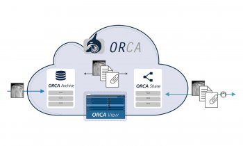
Among others, this has previously been affected by data protection restrictions between hospitals, but these are due to be lifted. Amongst technological innovations is nanotechnology, which provides new opportunities to guide light within the body using LED-illuminated instruments and a periscope with three cameras, which show round structures as flat. On the other hand, the professor does not give the controllable magnet capsule much of a chance – too much expense for too low a detection rate.
Finally, the latest news: Colon cancer might actually develop as a result of an infection.
Between 1981-85, Professor Christoph F Dietrich MD, now head physician at the Caritas Hospital in Bad Mergentheim, Germany, was a medical student at the Medizinische Hochschule in Hanover. In 1986-86, gastroenterology study followed in Seville, Spain, and at New York Medical College, USA. From 1988 to 89, following his German medical exam he received the ECFMG Certification, USA. He then specialised in Internal Medicine (1997), Gastroenterology and Hepatology (2000) and Pneumology (2002). In 1999 he joined Frankfurt University as an Assistant Professor and was appointed a full professor in 2005. He became a haematology and oncology specialist in 2008, in Palliative Care Medicine, and in Geriatric Medicine and Proctology in 2009 In 2010-12 he was a member of Frankfurt School of Finance & Management. For 2011-13 EFSUMB elected him President, and from 2012-13 he has been president of DGE-BV.
26.04.2013