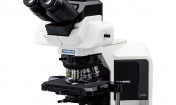
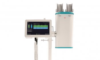
The new EVIS EXERA III
Major developments in endoscope technology were at the top of the agenda when European Hospital editors visited the European headquarters of Olympus in Hamburg, Germany.
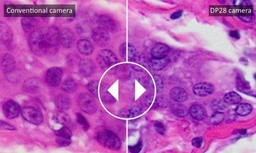
The EVIS EXERA III, a recent portfolio addition, makes significant contributions to quality and productivity in care, explained Olympus representatives John Cobain, Group Leader Gastroenterology, Business Unit Gastroenterology and Respiratory Endoscopy, and Mirko Feuring, Product Manager of Flexible Endoscopy for Germany, Austria and Switzerland. Image quality obviously plays a vital role in endoscopy. Thus, according to John Cobain, the new features developed for the EVIS EXERA III open up opportunities in diagnosis and treatment. Clearly detection is a key area, with the discovery of even the smallest lesions optimising patient care, and increased detection rates leading to reduced time spent on procedures. Characterisation is another important area. Supporting the physician in determining a diagnosis and treatment cuts the cost for pathology. Better images positively affect quality control and accelerated digitisation facilitates documentation.
It’s all about the image
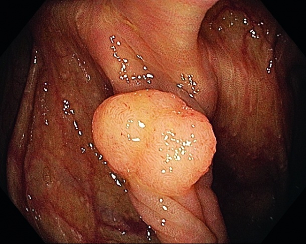
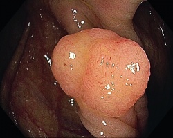
New image-related developments built into the EVIS EXERA III are dual focus, brighter Narrow Band Imaging (NBI), and pre-freeze. Dual focus is a two-stage optical technology that allows the user to select the position of the focal lens. The physician optimises the required depth of field to normal mode (5-100 mm / 170°) or near mode (2-6 mm, 160°). In normal mode, the highest resolution, wide field of view and the depth of field support detection, whereas in near mode the increased resolving power enhances in-situ characterisation. The depths of field of these modes overlap, facilitating their use. This, John Cobain emphasised, presents major advantages over zoom endoscopes. With the latter in tele mode, the depth of field is very limited and the field of view is also very narrow; wide and tele modes do not overlap and leave a wide area out of focus. EVIS EXERA III offers 1.5 brighter NBI – enabling twice the viewable distance when observing in NBI mode. The higher brightness of NBI will reduce the need to switch off NBI, and is bound to ‘unleash the true potential of NBI for detection’, Mirko Feuring pointed out. The pre-freeze function continuously stores acquired video footage; the least blurred frame is selected on demand.
Imaging is fundamental to characterisation
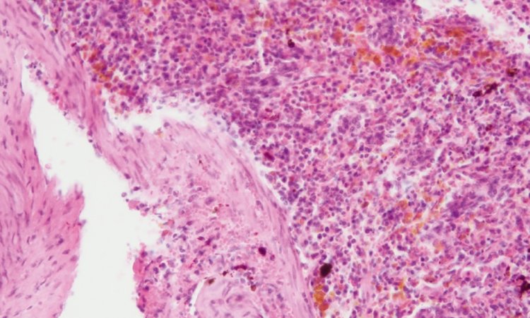
A number of attributes are crucial to optimal characterisation: high resolution near focus; high magnificationpositively affect quality control and accelerated digitisation facilitates documentation. levels without image deterioration; high contrast, enabling effective enhancement of vessels and pits. Key features of the EVIS EXERA III fulfil these requirements. Essential prerequisites for in-vivo characterisation are precise classification schemes; on this basis, invivo characterisation can provide faster diagnosis of pathology, and provides immediate feedback to the patient regarding surveillance.
Better control and optimised workflow
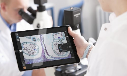
Responsive Insertion Technology (RIT) significantly improves endoscope handling. Passive bending integrates a passive, non-controllable bending section proximal to the active, controllable, distal bending section on PCF and CF scopes. This allows smooth passage through acute flexures or difficult anatomy. In contrast to standard scopes, High Force Transmission (HFT) efficiently transfers torque and lateral forces along the insertion tube, even when angulated. The endoscope comes with a new data management platform that supports staff in their work. Pictures in JPEG or TIFF format can be stored on USB media, allowing for review and comments. ‘Thanks also to IT connectivity for data sharing with clinical and administrative departments,’ added Mirko Feuring, ‘EVIS EXERA III is an interoperable, futureproof investment.’
04.09.2012