Optimised imaging challenges biopsy, but remains ‚ silver´ standard

“The powerful capabilities for thoracic imaging can often be sufficient to make a diagnosis to treat the patient,” notes Philippe Grenier, MD. If CT findings, for example, are typical for idiopathic pulmonary fibrosis, there should no longer a need for a biopsy, which is good for the patient.

Philippe A Grenier
Philippe A Grenier

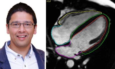
A former President of ESTI, he will present, during a session dedicated to Interstitial Lung Disease, a review of high-resolution CT techniques with an emphasis on “Optimising the Image.” “Imaging has become a fantastic tool for identifying the diseases,” he said, “but remains a “silver standard” for assessment rather than a definitive gold standard for diagnosis.”

“It requires a multi-disciplinary approach to arrive at the level of confidence that can be considered golden,” said Grenier, whose cross-discipline experience includes serving as the Head of the Medical Board for the Pitié-Salpêtrière Hospital in Paris. Professor of Radiology at Pierre & Marie Curie University, Paris, Grenier demonstrated the effectiveness of combining CT findings with clinical and chest radiographic assessments in 1994. The paper published in Radiology showed that a correct diagnosis based on clinical data was obtained in 29% of cases, that radiography was correct in only 9%, and that CT correctly identified the condition in 36% of cases. When the three disciplines were combined, diagnostic sensitivity rose to 80%.

Returning to his example, however, Grenier said that where the combined assessments do not identify a cause for the symptoms, or the findings for an idiopathic pulmonary disease are equivocal, then a biopsy is required. “Lung biopsy cannot be considered a gold standard either,” he said. “There is interobserver variability between pathologists, as there will be between radiologists for interpreting HRCT findings.”Improvements to CT capabilities mean that imaging will continue to enhance its position for diagnostic confidence and, thereby, help reduce the need for the invasive biopsy procedure.

“Today it is possible in a single breath-hold to acquire volumetric data of the entire lung, which through post-processing techniques give us the possibility of a better analysis of the relationship between peripheral airways and the lung periphery, to distinguish the difference between honeycombing or traction bronchiolectasis,” he said. “We have many possibilities to improve the information coming from imaging.”
 
Volumetric acquisitions are justified in providing the whole lung assessment and for their value in following the patient after treatment, as it is easier to have equivalent examinations to compare with the same level as the previousexamination. “The problem is we are delivering higher dose than we did with HRCT,” he said. This discussion will be a central point of his presentation on optimising imaging for interstitial lung disease. He will demonstrate that with iterative reconstruction it is possible to have equivalent results. “This is my objective,” he said, “to say that if you do low-dose volumetric assessment with post processing techniques, you will have the same result.”
 
Profile
 
Philippe A Grenier is Professor and Chairman of Radiology (General Diagnosis) and heads the Medical Board at Pitié-Salpêtrière Hospital at the Pierre & Marie Curie University in Paris, France. Professor Grenier was a founding member of the European Society of Thoracic Imaging and a member of leading societies, such as the European Society of Radiology and the International Society of Strategic Studies in Radiology. He is also an Honorary Member of many international radiological societies, e.g. in North America, Canada, Sweden, Italy, Japan, Austria, and Argentina; in 2007, he earned the Gold Medal of European Congress and Association of Radiology.
 
With main interests in chest imaging, lung cancer, interstitial lung disease, airway disease, COPD and asthma, his scientific publications are notable. He not only serves on the editorial boards of European Radiology , Academic Radiology, Journal of Thoracic Imaging and is on the Advisory Board of Acta Radiologica, but he also authored numerous peer reviews, book chapters and books, and has presented 155 guest lectures.


19.06.2012

More on the subject:

Related articles

Photo

News • OCT and IR-SLO imaging

Eye scans for early detection of Multiple Sclerosis

Doctors may soon have a new, high-tech way to diagnose Multiple Sclerosis using special eye scans. This could make it easier and faster for people to find out if they have MS.

Photo

Article • Delivering more efficient healthcare

How teleoperation is changing radiology

As opportunities for teleoperations rapidly expand within radiology, the concept is being deployed across an array of modalities to deliver more efficient healthcare. A range of speakers covered the…

Photo

News • Cardiac imaging analysis

AI greatly speeds up heart chamber segmentation from MRI scans

From 45 minutes to just a few seconds: A new computer model utilises AI to examine cardiac MRI scans in the four-chamber plane, potentially offering speedy and dependable heart health evaluation.

Related products

Subscribe to Newsletter