Credit: Paulina Wyszkiewicz
News • Targeted treatment
Lung-imaging shows cause of long COVID symptoms
Many who experience what is now called "long COVID" report feeling brain fog, breathless, fatigued and limited in doing everyday things, often lasting weeks and months post-infection. Using functional MRI with inhaled xenon gas, researchers have now identified for the first time that these debilitating symptoms are related to microscopic abnormalities that affect how oxygen is exchanged from the lungs to the red blood cells.
The LIVECOVIDFREE study, based at five centers across Ontario, and led by Western University professor Grace Parraga, is the largest MRI study of patients with long COVID. The research is the first to show a potential cause of these long COVID symptoms. By understanding the cause, team members responsible for patient care have been able to target treatment for these patients.
"I think it is always a conundrum when someone has symptoms, but you can't identify the problem. Because if you can't identify the problem, you can't identify solutions," said Parraga, Tier 1 Canada Research Chair in Lung Imaging to Transform Outcomes at Western's Schulich School of Medicine & Dentistry.
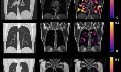
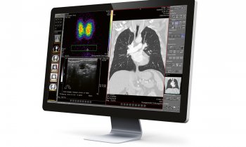
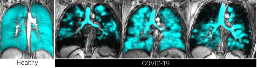
By having study participants inhale polarized xenon gas while inside the MRI, the researchers see in real-time the function of the 300-500 million tiny alveolar sacs, which are about 1/5 of a mm in diameter and responsible for delivering oxygen to the blood.
"With our MRI technique, we can watch in real time the air moving through the alveolar membrane and through to the blood cells; and we can actually see the function of the tiny alveolar sacs in the lungs," said Parraga. "What we saw on the MRI was that the transition of the oxygen into the red blood cells was depressed in these symptomatic patients who had had COVID-19, compared to healthy volunteers."
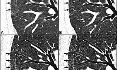
Further CT scans pointed to "abnormal trimming" of the vascular tree, indicating an impact on the tiny blood vessels that deliver red blood cells to the alveoli to be oxygenated. There also doesn't appear to be any difference in severity of this abnormality between patients who had been hospitalized with COVID-19, and those who recovered without hospitalization, the study said. This is an important finding as the latest wave of COVID-19 infection has affected large numbers of people who did not need hospital-based care.
"For those who are symptomatic post-COVID, even if they hadn't had a severe enough infection to be hospitalized, we are seeing this abnormality in the exchange of oxygen across the alveolar membrane into the red blood cells," said Parraga.
The researchers recruited patients with suspected long COVID from two hospitals in London, Ontario: London Health Sciences Centre's (LHSC) Urgent COVID-19 Care Clinic (LUC3), and St. Joseph's Health Care London's post-acute COVID-19 program. Participants were those with persistent shortness of breath more than six-weeks post-infection. Some study participants were still symptomatic after 35 weeks.
Study co-author Dr. Michael Nicholson is a respirologist with St. Joseph's post-acute COVID-19 program, former member of the LUC3 clinical team at LHSC, and an associate scientist at Lawson Health Research Institute. He said patients who were describing these symptoms were also showing normal results on clinical breathing tests.
Credit: Alexander Matheson
"We were looking for further modalities to look at their lung function that were not found through traditional clinical testing," said Nicholson. "The findings allowed us to show that there was a physiological impact on their lungs that correlated with their symptoms."
Study participant Alex Kopacz described his experience with COVID-19 as "harrowing." He was admitted to LHSC's University Hospital with the virus in 2021. A young, fit Canadian bobsledder and Olympic gold medalist, he never imagined that he would still be struggling to breathe months after infection.
"I was on oxygen for almost two months after COVID, and it took me almost three months to get to a place where I could go for a walk without gasping for air," said Kopacz. "The take-home message for me is that we have to remember that this virus can have very serious long-term consequences, which are not trivial. In my case, prior to getting sick, I didn't think it would really affect me."
A one-year follow-up is now underway to better understand these results longitudinally. The study was done in collaboration with researchers at LHSC, St. Joseph's, Lakehead University, McMaster University, Toronto Metropolitan University and Sick Kids Hospital in Toronto.
The research was published in Radiology.
Source: University of Western Ontario
29.06.2022