News • Zooming in
Breast cancer map reveals how mutations shape the tumour landscape
Scientists have created one of the most detailed maps of breast cancer ever achieved, revealing how genetic changes shape the physical tumour landscape.
An international team of scientists, brought together by a £20 million Grand Challenge award from Cancer Research UK, has developed intricate maps of breast tumour samples, with a resolution smaller than a single cell. These maps show how the complex cancer landscape, made up of cancer cells, immune cells and connective tissue, varies between and within tumours, depending on their genetic makeup. This technique could one day provide doctors with an unparalleled wealth of information about each patient’s tumour upon diagnosis, allowing them to match each patient with the best course of treatment for them.
The researchers now published their findings in Nature Cancer.
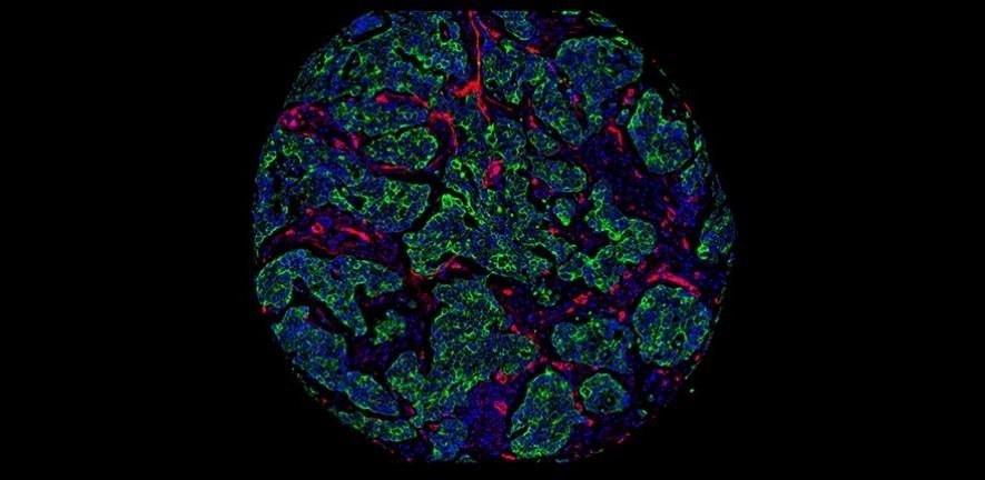
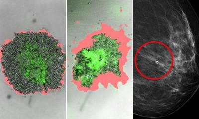
Credit: H. Raza Ali
In the future, it could also be used to analyse tumours during treatment, allowing doctors to see in unprecedented detail how tumours are responding to drugs or radiotherapy. They could then modify treatments accordingly, to give each patient the best chance of beating the disease. Dr Raza Ali, lead author of the study and junior group leader at the Cancer Research UK Cambridge Institute, said: “At the moment, doctors only look for a few key markers to understand what type of breast cancer someone has. But as we enter an era of personalised medicine, the more information we have about a patient’s tumour, the more targeted and effective we can make their treatment.”
The researchers studied 483 different tumour samples, collected as part of the Cancer Research UK funded METABRIC study, a project that has already revolutionised our understanding of the disease by revealing that there are at least 11 different subtypes of breast cancer. The team looked within the samples for the presence of 37 key proteins, indicative of the characteristics and behaviour of cancer cells. Using a technique called imaging mass cytometry, they produced detailed images, which revealed precisely how each of the 37 proteins were distributed across the tumour.
The researchers then combined this information with vast amounts of genetic data from each patient’s sample to further enhance the image resolution. This is the first time imaging mass cytometry has been paired with genomic data. These tumour ‘blueprints’ expose the distribution of different types of cells, their individual characteristics and the interactions between them. By matching these pictures of tumours to clinical information from each patient, the team also found that the technique could be used to predict how someone’s cancer might progress and respond to different treatments.
This team is making incredible advances, helping us to peer into a future when breast cancer treatments are truly personalised
David Scott
Professor Carlos Caldas, co-author of the study from the Cancer Research UK Cambridge Institute, said: “We’ve shown that the effects of mutations in cancer are far more wide-ranging than first thought. They affect how cancer cells interact with their neighbours and other types of cell, influencing the entire structure of the tumour.”
The research was funded by Cancer Research UK’s Grand Challenge initiative. By providing international, multidisciplinary teams with £20 million grants, this initiative aims to solve the biggest challenges in cancer.
Dr David Scott, director of Grand Challenge at Cancer Research UK said: “This team is making incredible advances, helping us to peer into a future when breast cancer treatments are truly personalised. There’s still a long way to go before this technology reaches patients, but with further research and clinical trials, we hope to unlock its powerful potential.”
Source: © University of Cambridge (CC BY 4.0)
18.02.2020